Chemoenzymatic Synthesis of Purine Modified 3′-deoxy-D-ribonucleosides and Evaluation of Their Antiproliferative Activity
Abstract
Chemoenzymatic synthesis of a series of purine modified 3′ deoxyribonucleosides was studied from purine derivatives as acceptors and an excess of 3′-deoxyuridine as the donor, prepared by the chemical deamination of 3′-deoxycytidine, using recombinant uridine and purine nucleoside phosphorylases as biocatalysts in enzymatic reactions. Eight purine nucleosides were prepared in 24%-84% yields after column chromatography. Among the tested purine derivatives, 2,6-dichloropurine was found to be the best substrate in the enzymatic transglycosylation reaction catalyzed by the E. coli purine phosphorylase (PNP) via intermediate 1-phosphate-3-deoxy-D-ribofuranose, and dihalogenated purine 3'-deoxyriboside was prepared in 84% yield. The anticancer nucleoside, cordycepin, was synthesized by the enzymatic transglycosylation reaction of adenine in 70% yield from 3´-deoxyuridine. Two enzymatic approaches to 2-fluorocordycepin were tested from 2-fluoroadenine or 2-fluoroadenosine and 3´-deoxyuridine as the donor of 3-deoxy-D-ribofuranose in the transglycosylation of the 2-fluoropurine using the recombinant PNP. 3′-Deoxyribofuranosides of 2,6-chloro- and 6-chloro-purine were utilized as starting compounds for preparing novel purine modified nucleosides by the nucleophilic substitution reactions of the chlorine atom with cyclic amines or acylation reaction. A series of modified purine 3′-deoxyribonucleosides were evaluated for their in vitro antiproliferative activity on leukemia cell lines HL-60 and K-562.
Article Information
- Received
- Accepted
- Published
Academic Editor: Loai Aljerf, Department of Life Sciences, Faculty of Dentistry, University of Damacus.
Checked for plagiarism: Yes
Review by: Single-blind
Copyright © 2026 Darya V. Kozlovich, et al.
 This is an open-access article distributed under the terms of the Creative Commons Attribution License, which permits unrestricted use, distribution, and reproduction in any medium, provided the original author and source are credited.
Corresponding author: Grigorii G. Sivets, Institute of Bioorganic Chemistry, National Academy of Sciences, 220084 Minsk, Acad. Kuprevicha 5/2, Belarus —
Competing Interests
The authors have declared that no competing interests exist.
Funding
No specific funding statement was provided by the authors.
Data Availability
No data-availability statement was provided by the authors.
Acknowledgements
This study was supported by a grant from FOI «Chemical processes and technologies», s/p «Chemical foundations of life activity processes» (Grant 2.3.04). The authors express gratitude to researchers, Institute of Bioorganic Chemistry of NAN of Belarus, for obtaining NMR spectral data, P.C. Shabunya for providing mass-spectral data, are grateful to O.B. Panibrat, S.E. Ogurtsova for their kind assistance in biological assays.
Citation:
Introduction
Among a great diversity of bioactive purine and pyrimidine nucleosides, 3′-deoxy-ribonucleosides display antibacterial, antitrypanosomal and anticancer activities 1, 2, 3, 4. A wide spectrum of their interesting biological properties is due to the lack of a 3′-hydroxy group and formation of nucleoside 5′-O-triphosphates that lead to the inhibition of the process of RNA and DNA synthesis. Discovery of anticancer activity and research of mechanisms of 3´-deoxyadenosine (cordycepin), a natural adenosine analogue of fungal origin, in inhibiting cancer cells has stimulated considerable interest to chemistry and biology of this type of deoxynucleosides (Figure 1) 5. Recently, it was found that a new metabolic pathway for therapeutic effect of 3′-deoxyadenosine can proceed via generation of 3′-deoxyinosine as the main metabolite followed by its conversion to cordycepin 5´-O-triphosphate 6.
3′-Deoxyanalogues of cordycepin containing a fluorine or chlorine atom at the C-2 carbon atom of the purine as well as 3´-deoxyribofuranosides of 7-arylsubstituted 7-deazapurine derivatives are of particular interest due to their potent in vitro activities against Trypanosoma brucei and cruzi (Figure 1) 3, 7. Besides, 3´-deoxyadenosine has recently been shown to have capability to potently inhibit in vitro the multiplication of the new resistant strains of coronavirus (SARS-CoV-2) in comparison with remdesivir as the reference drug 8.
Figure 1. Biologically active purine 3′-deoxyribonucleosides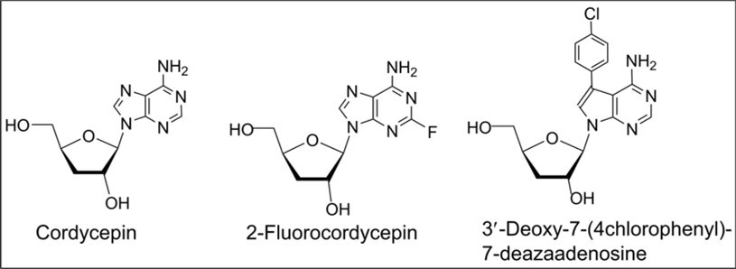
Download figure
Chemical synthesis of 3′-deoxyribonucleosides has been explored using the two main approaches: (i) the introduction of protective groups in the carbohydrate moiety of natural pyrimidine and purine ribonucleosides, the preparation of intermediate 3′-O-thiocarbonyl (PTC) nucleoside derivative or 3′-halogenated nucleosides followed by their radical deoxygenation or catalytic debromination, and deprotection (Scheme 1, a and b) 9, 10, 11 and (ii) the convergent synthesis of 3′-deoxyribonucleosides by the glycosylation reactions of various heterocyclic bases with acyl protected 3-deoxy-D-ribofuranose derivatives 1, 3, 12, 13 as the key intermediates prepared by multistep procedures from D-xylose or D-glucose (Scheme 1, c). Chemical modifications of available natural nucleoside, or the coupling reactions of monosaccharide derivatives with heterocyclic bases resulted in the target nucleosides by stereoselective or regioselective syntheses with well-known shortcomings, such as a multistep process, the application of various protective groups for the introduction in the heterobase and sugar followed by their removal after the radical, catalytic deoxygenation steps or the condensation reactions.
Scheme 1. Previous chemical syntheses of pyrimidine and purine 3'-deoxyribonucleosides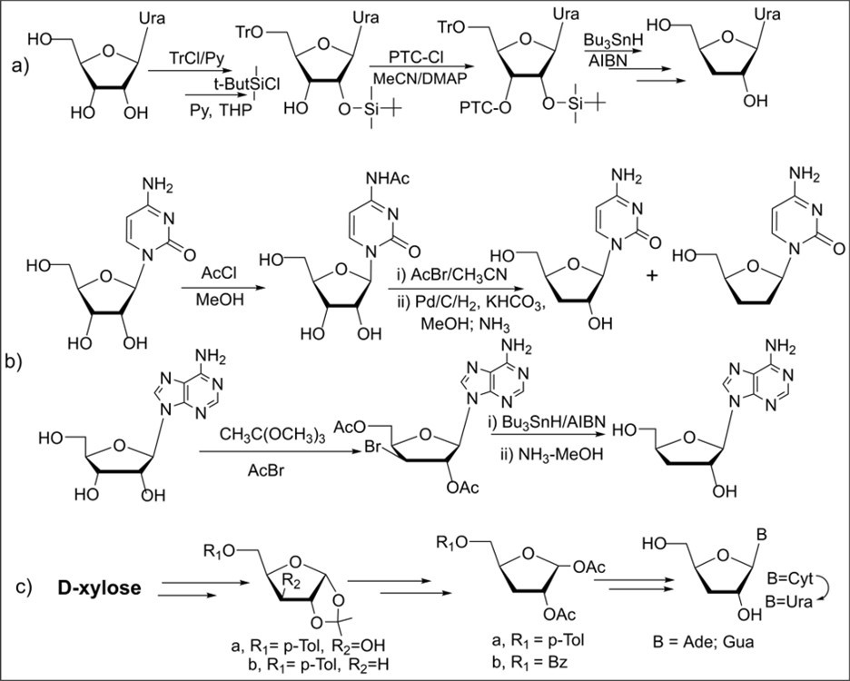
Download figure
By contrast, in an enzymatic approach, nucleoside phosphorylases 14, 15, 16, 17 can be used as biocatalysts for efficient preparation of purine deoxy, fluorodeoxy nucleoside analogues and 3′-deoxyribonucleosides via the enzymatic transglycosylation reactions 18, 19. The chemoenzymatic synthesis of 3′-deoxy-β-D-ribofuranosyl purines has earlier been reported starting from 2,6-diaminopurine as an acceptor and 3′-deoxycytidine (3′dCyd) as a donor of 3-deoxy-D-ribofuranose using the selected intact E. coli BM-11 and BMT-4D/1A cells as a biocatalyst (Scheme 2, a) 4.
During the studied enzymatic reaction 3′dCyd is deaminated to 3′-deoxyuridine by the powerful cytidine deaminase of E. coli BM-11 cells to give the intermediate 3-deoxy-D-ribofuranose-1-phosphate after the phosphorolysis reaction of the latter and the purine nucleoside via transglycosylation. The 3′-deoxyriboside of 2,6-diaminopurine was used to prepare related 3′-deoxyguanosine and 2-fluorocordycepin through the chemical transformations in the purine heterobase.
2-Fluoro- or chlorocordycepins possessing antibacterial activities 7 were also produced using 3′-deoxyribofuranosides of adenine or hypoxanthine as sugar donors in the presence of the recombinant E. coli PNP as a single biocatalyst (Scheme 2, b). The transglycosylation reaction of 2-fluoroadenine catalyzed by PNP gave 2-fluorocordycepin at 40 0C in 62% yield using 3′-dAdo as a donor of the 3-deoxy-D-ribofuranose (Scheme 2) 18. 3′-Deoxyinosine prepared in four steps from commercially available inosine was utilized in the enzymatic synthesis of 2-halogenated cordycepin derivatives (Scheme 2). 3′-Deoxyadenosine and 3′-deoxyinosine were found to be satisfactory donors of 3-deoxy-D-ribofuranose in the E.coli PNP catalyzed the transglycosylation reaction of 2-fluoro- or 2-chloroadenine. Using two enzymatic approaches the synthesis of 2-fluorocordycepin has been studied and its anticancer activity in vitro has recently been described by Arnautova et al.19. It was shown that the fluoro-containing cordycepin derivative exhibits the cytotoxic potential against a number of cancer cell lines (Jurkat, Raji, MCF-7, THP-1, U937, A549, LS174T).
Scheme 2. Previous enzymatic syntheses of purine 3'-deoxyribonucleosides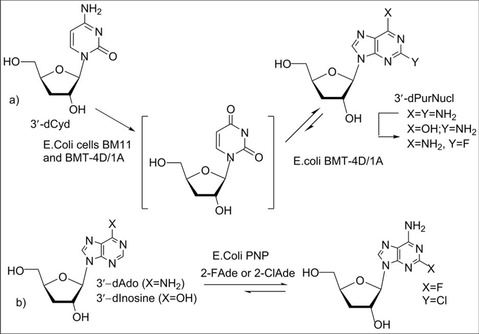
Download figure
Enzymatic syntheses of deoxy and fluorodeoxy nucleoside analogues were previously described through enzyme-catalyzed transglycosylation reactions involving recombinant pyrimidine and purine nucleoside phosphorylases as biocatalysts 20, 21. Substrate specificity of several pyrimidine nucleoside phosphorylases 21 has been studied towards pyrimidine 2′-deoxy, 3′-fluoro-3′-deoxy and 3′-deoxyribonucleosides. 2′ and 3′-Deoxyuridines were shown to possess ability to undergo efficient phosphorolysis reaction with the mutant pyrimidine phosphorylase of Thermus thermophilis or recombinant uridine phosphorylase of Escherichia coli and may be used as donors of the sugar moiety in the tandem enzymatic synthesis of purine nucleosides. Besides, recombinant nucleoside phosphorylases from E.coli were used for the synthesis of pharmaceutically valuable nucleosides 22. In the present work, we report stereoselective chemoenzymatic synthesis of a series of purine modified 3´-deoxyribonucleosides using recombinant nucleoside phosphorylases starting from 3´-deoxyuridine, preparation of N-6-substituted purine 3′-deoxyribonucleosides by chemical modification of halogenated purine nucleoside derivatives and evaluation of their antiproliferative activity.
Results and discussion
3´-Deoxy-D-cytidine was prepared from cytidine by the known method, elaborated earlier for L-enantiomeric nucleosides - β-L-ddCyd and β-L-3´dCyd 10, 23, and used to synthesize 3´-deoxyuridine as the starting compound for enzymatic studies. Several multistep approaches have been investigated for preparing 3´-deoxyuridine from D-xylose 12, 24 and uridine 9, 12, 25. Deamination reactions of purine and cytosine nucleosides or nucleotides, their kinetics have earlier been studied with nitrous acid 26, 27, 28. Synthetic deamination procedure to prepare 3´-deoxyuridine by nitrite-assisted reaction of 3´-deoxycytidine has not been described in the literature. We investigated deamination of 3´-deoxycytidine (1) with sodium nitrite in acetic acid under various conditions. 3´-Deoxyuridine (3), as the key nucleoside for enzymatic reactions, was prepared by the treatment of the cytosine nucleoside with excess of NaNO2 in glacial CH3COOH at room temperature in 40% yield along with its 5´-O-acetyl derivative 2 (5%) after column chromatography on silica gel (Scheme 3).
A more efficient method for nitrite-assisted deamination of 1 was explored using excess of sodium nitrite in 50% aq acetic acid at room temperature followed by the mild heating and treatment of the reaction mixture with aqueous ammonia. In this case, 3´-deoxyuridine (3) was synthesized by deamination of 1 with nitrous acid in 52% yield. However, tested reaction conditions afforded less yield of 3´-deoxyuridine than the methods described previously via deamination reactions of 3´-deoxycytidine or the 3´,5´-di-O-acylated nucleoside derivative, prepared from D-xylose, with sodium bisulfite or nitrite, respectively 24, 29.
Preceding studies of phosphorolysis reactions of 2´- and 3´-deoxyuridines catalyzed by the recombinant E.coli uridine phosphorylase, mutant pyrimidine phosphorylase of Thermus thermophilisin phosphate buffer showed that degree of phosphorolysis for 3´-deoxyuridine was greatly lower than that of 2´-deoxyuridine under similar conditions with E.coli UP, but efficient phosphorolysis reactions were observed for the both nucleosides with mutPyrNPase from T. thermophilis21. Synthesis of 2-halogenated 3´-deoxyadenosine derivatives was earlier developed from purine 3´-deoxyribonucleosides 18 or individual 1-phosphate-3-deoxy-D-ribofuranose 19 and optimal conversion of the purine base into the nucleoside was shown to proceed in the presence of excess of E.coli PNP (4000-4550 units or more 1000 units per 1 mmol of purine base). Based on previous results on study of the phosphorolysis reaction of 3´-deoxyuridine with E.coli UP, properties of the given recombinant enzyme 22 and the findings on syntheses of 2-halogenated cordycepins, testing of optimal reaction conditions at small scale for enzymatic synthesis of purine 3´-deoxyribonucleosides from 6-chloropurine or 6-chloro-2-aminopurine in 10 mM potassium phosphate (pH 7.4) was carried out under mild heating varying amounts of E.coli UP and PNP phosphorylases 30, 31 and reaction time. It was found that corresponding purine modified nucleosides can be prepared in good yields from the purine bases and excess of donor (1:2), 3´-deoxyuridine, in the presence of excess of UP (3900 - 5900 units per 1 mmol) and PNP (>4900 units per 0.5 mmol of base) in 10 mM potassium phosphate buffer under mild heating.
Enzymatic synthesis of a series of purine modified 3´-deoxyribonucleosides 5-12 was carried out in 10 mM potassium phosphate (pH 7.4) under mild heating using recombinant E.coli UP and PNP phosphorylases 22 as biocatalysts and 3´-deoxyuridine as the donor of 3-deoxyribose moiety (Scheme 3, Table 1). The target purine 3´-deoxyribonucleosides were prepared from purine bases via the tandem transglycosylation reactions in the presence of excess of recombinant enzymes and isolated by column chromatography on silica gel in 24%-84% yields (experimental part). Besides, the substrate specificity of E.coli PNP phosphorylase for different modified purine heterocyclic bases was explored in enzymatic reactions for preparation of purine modified 3´-deoxyribonucleosides (Table 1). Among 2,6-halogenated purine derivatives with chlorine or fluorine atoms, 2,6-chloropurine revealed the best substrate properties in the enzymatic reaction with intermediate 1-phosphate-3-deoxy-D-ribofuranose 4 catalyzed by recombinant purine phosphorylase from E.coli and the corresponding 3´-deoxyribonucleoside 5 was synthesized in a high yield (86%) from 3´-deoxyuridine 3. Using 2-chloroadenine as an acceptor in the transglycosylation reaction under consideration catalyzed by PNP, 2-chloro-3′-deoxyadenosine 11 was synthesized in 24% yield after column chromatography. It was also found that 6-chloro and 6-chloro-2-fluoro-purines can be used in tandem enzymatic syntheses to prepare purine modified nucleosides 6 and 7, which were isolated in 73% and 32% yield, respectively. The application of purine derivatives with amino group at 6 or 2-position of the heterocycle in enzymatic reactions (Scheme 3) lead to corresponding 3´-deoxyribonucleosides in moderate yields. Among modified purines tested in enzymatic transglycosylation, 2-amino-6-chloropurine gave good yield of purine 3´-deoxyribonucleoside 8 (69%). Adenine was also found to be a good substrate for PNP and cordycepin 10 was prepared in a high yield (70%) from 3´-deoxyuridine (3) after the enzymatic reaction followed by column chromatography on silica gel.
Scheme 3. Synthesis of 3'-deoxyuridine via the deamination reaction and purine modified 3'-deoxyribonucleosides using the enzymatic transglycosylation reactions. Reagents and conditions. a1) 1, NaNO2/CH3COOH, rt, 96 h, 2 (5%) and 3 (40%); a2) 1, NaNO2/50% aq.CH3COOH, rt, 24 h, then 40 0C, 30 min, 3 (52%); b) 2,6-disubstituted purine, 3′-deoxyuridine (3) (mol ratio 1:1.0-2.0), 10 mM potassium phosphate buffer (pH 7.4), UP and PNP E.coli, 40 0C, 48-72 h, purine 3'-deoxyribonucleosides 5-12, 24%-84%.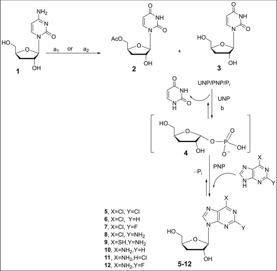
Download figure
Enzymatic synthesis was tested from 5′-O-acetylated 3′-deoxyuridine 2 as a donor of the carbohydrate moiety to prepare 2,6-chloropurine 3′-deoxyribonucleoside derivative under conditions similar to the efficient preparation of the 2,6-chloropurine nucleoside 5 catalyzed by E.coli PNP (Table 1, entry 1). In this case, formation of 5'-O-acetylated 3'-deoxyribonucleoside of 2,6-chloropurine was not observed via phosphorolysis reaction and the subsequent base exchange reaction in the presence of PNP according to 1H NMR analysis (CDCl3) of the reaction mixture after studying conversions of the starting O-acylated nucleoside and 2,6-chloropurine.
Table 1. Enzymatic synthesis of 3´-deoxyribonucleosides 5-12 from 3′-deoxyuridine using NPs1| Entry | Purine base | (Donor 3′-dUrd)(mM) | Acceptor (Purine heterobase)(mM) | Synthesizednucleoside | Time, h | 3′-deoxynucleoside number and yield (%)2 |
| 1 | 2,6-diClPur | 1.2 | 0.61 | 2,6-diClPur-3′-dR | 72 | 5 (84) |
| 2 | 6ClPur | 0.97 | 0.48 | 6-ClPur-3′-dR | 72 | 6 (73) |
| 3 | 2F,6Cl-Pur | 0.2 | 0.2 | 2-F,6-Cl-Pur-3′-dR | 48 | 7 (32) |
| 4 | 2NH2,6Cl-Pur | 0.24 | 0.12 | 2-NH2,6-Cl-Pur-3′dR | 72 | 8 (69) |
| 5 | 2NH2,6SH-Pur | 0.3 | 0.15 | 2-NH2,6-SH-Pur-3′-dR | 72 | 9(30) |
| 6 | Ade | 0.74 | 0.37 | 3′-dAdo | 72 | 10 (70) |
| 7 | 2-ClAde | 0.13 | 0.11 | 2Cl-3′-dAdo | 48 | 11 (24) |
| 8 | 2-FAde | 0.44 | 0.22 | 2F-3′-dAdo | 72 | 12 (40) |
Synthesis of 2-fluorocordycepin 12 was also explored using 2-fluoroadenosine 1332, 33, a donor of 2-fluoroadenine, and 3′-deoxyuridine as that of 3-deoxy-D-ribofuranose-1-phosphate in the cross-glycosylation reaction in the presence of two recombinant nucleoside phosphorylases (Scheme 4). Conversion of 2-fluoroadenosine 13 into 2-fluorocordycepin 12 was investigated in the presence of two biocatalysts for 72 h (monitored by TLC - ethyl acetate-EtOH-H2O – 7:1:0.5) under mild heating (40 0C) with the use of 1.8 molar excess of 3′-deoxyuridine. After completing enzymatic synthesis and treatment of the reaction mixture with methanol (experimental part), 1H NMR analysis showed the presence of uracil, 3′-deoxyuridine and the target 2-fluorocordycepin as the main components in a ratio of 0.7:1.6:0.85 along with 2F-Ade as minor component according to 19F NMR spectrum in DMSO-d6 (two singlets at 53.2 ppm and 54.7 ppm, signals of F-2 nuclei for nucleoside 12 and 2-fluoropurine, respectively). The target nucleoside was prepared in 57% yield after column chromatography on silica gel.
Scheme 4. Chemoenzymatic synthesis of 2-fluorocordycepin from 2-fluoroadenosine and 3'-deoxyuridine. Reagents and conditions. 2-Fluoroadenosine (13), 3′-deoxyuridine (3) (mol. ratio 1:1.82), 5 mM phosphate buffer (pH 7.2), UP E.coli (5600 units) and PNP E.coli (7000 units), 40 0C, 72 h, CC on silica gel, 2-fluorocordycepin (12), 57%.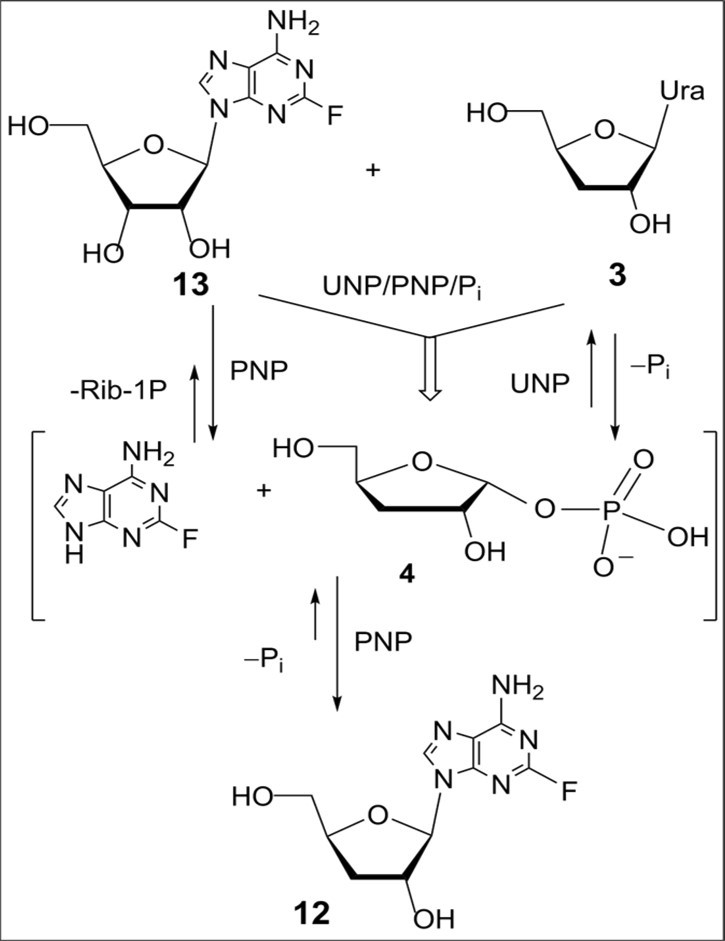
Download figure
Thus, two approaches towards the biologically important 2-fluoropurine nucleoside derivative based on three- or two-step enzymatic conversions were studied using the excess of the starting 3'-deoxyuridine. The enzymatic cross-glycosylation between two nucleosides (2-fluoroadenosine and 3′-dUrd) gave higher yield of nucleoside 12 than that of the enzymatic approach studied through intermediate formation of 1-phosphate of 3-deoxy-D-ribofuranose followed by the coupling with 2-fluoroadenine catalysed by E.coli PNP (Table 1, entry 8). A low solubility of 2F-Ade in the water reaction mixtures is the limiting factor for tested approaches. Structures of synthesized nucleosides were characterized by NMR spectral data and mass-spectroscopy.
Next, the chemoenzymatic approach explored for a series of purine 3′-deoxyribonucleosides from 3′-deoxyuridine makes it accessible 3′-deoxyribofuranosides of 2,6-di and 6-monohalogenated purines for further synthesis of new purine nucleosides with potential biological activity.
With 3′-deoxy-D-ribofuranosides of 2,6-chloro- (5) and 6-chloro-purine (6) in our hands, preparation of novel N6-substituted cordycepin analogues was carried out by the nucleophilic substitution reactions of the chlorine atom with N-containing nucleophilic agents (cyclic amines) at C6 position of the purine heterocycle or nucleosides modified in the carbohydrate moiety via acylation reactions (Scheme 5). The treatment of nucleosides 5 and 6 with excess of heterocyclic amine such as piperidine in anhydrous ethanol in the presence of EtNiPr2 gave N6-substituted purine 3′-deoxynucleosides 14 and 17 in 98% yields. The reaction of 6-chloropurine nucleoside 6 with pyrrolidine in acetonitrile resulted in N-6-pyrrolidinyl derivative of nucleoside 18 with 90% yield after column chromatography on silica gel. Acylation of 2,6-dichloropurine 3′-deoxyribonucleoside 5 with isobutyric anhydride in acetonitrile at room temperature in the presence of EtNiPr2 gave a mixture of acylated nucleosides 15 and 16 which was separated by column chromatography on silica gel. 3′,5′-Di-O-isobutiroyl-3′-deoxy-β-D-ribofuranosyl 2,6-dichloropurine 15 and 2′-O-acylated nucleoside 16 were isolated in 15% and 36% yields, respectively. Reaction of 6-chloropurine nucleoside 6 in the presence of EtNiPr2 with cyclic amino acid such as L/D-proline methyl ether of hydrochloride in acetonitrile at room temperature gave a mixture of diastereomeric N6-proline substituted nucleosides 19 (a d/r ratio – 1:0.91 according to 1H NMR spectrum, signals of H-8 and H-2 protons) with the proline residue at C6 position of the purine, which was isolated by column chromatography on silica gel in 78% yield (Scheme 5). Attempts to separate a mixture of nucleosides 19 by TLC or column chromatography on silica gel were unsuccessful under various tested conditions. Structures of new purine 3′-deoxyribonucleosides 14-19 modified in the heterobase and carbohydrate moiety were supported by NMR spectral data and masspectroscopy. The presence of the CH2 groups of N6-pyrrolidinyl and piperidinyl substituents in the purine nucleosides 14,17, and 18 was confirmed by the 1H and 13C NMR spectra. The 1H NMR spectra of N6, N6-penta- and tetramethylene-3′-deoxyadenosine derivatives contained resonance signals of protons for -N-CH2 fragments and the CH2 groups in two fields at 3.0-4.1 ppm and 1.5-2.0 ppm, respectively. The 13C spectra of the N6-cyclic amine-substituted nucleosides 14, 17, 18 and 19 contained resonance signals of the CH2 groups and C-3′ in the form of singlets at 24.2 - 48.0 ppm and 33.0 - 39.0 ppm, respectively. 1H NMR spectra for nucleosides 15 and 16 acylated in the carbohydrate moiety show the presence of resonance signals for protons of isobutyroyl protective groups in the form of multiplets at 1.5-1.6 ppm and 2.6-2.66 ppm. Besides, the assigned structures of di- and mono-O-acylated nucleosides of 2,6-dichloropurine were also confirmed by comparison of 13C spectral data, chemical shifts for carbon atoms of the 3-deoxy-D-ribofuranose moiety, with those of unprotected 2,6-dichloropurine 3′-deoxy-D-ribofuranoside 5. In 19F spectra of 2-fluoropurine nucleosides 7 and 12, signals for the 19F nuclei displayed as singlets at 51.96 and 53.2 ppm.
Scheme 5. Synthesis of purine 3'-deoxyribonucleosides modified in the heterobase and carbohydrate moiety from chlorinated purine nucleosides 5 and 6. Reagents and conditions. a1) 5, ahydr. EtOH, piperidine, DIEPA, rt, 2h, 85 0C, 14, 98%; b) 5, (i-BuCO)2O, CH3CN, DIEPA, rt, 20h, 15, 15%, 16, 36%; a2) 6, ahydr. EtOH, piperidine, DIEPA, rt, 2h, 85 0C, 17, 98%; c) 6, ahydr. EtOH, pyrrolidine, Et3N, rt, 2h, 85 0C, 18, 97%; d) 6, CH3CN, D/L-proline methyl ether hydrochloride, DIEPA, rt, 48h, 19, 92%.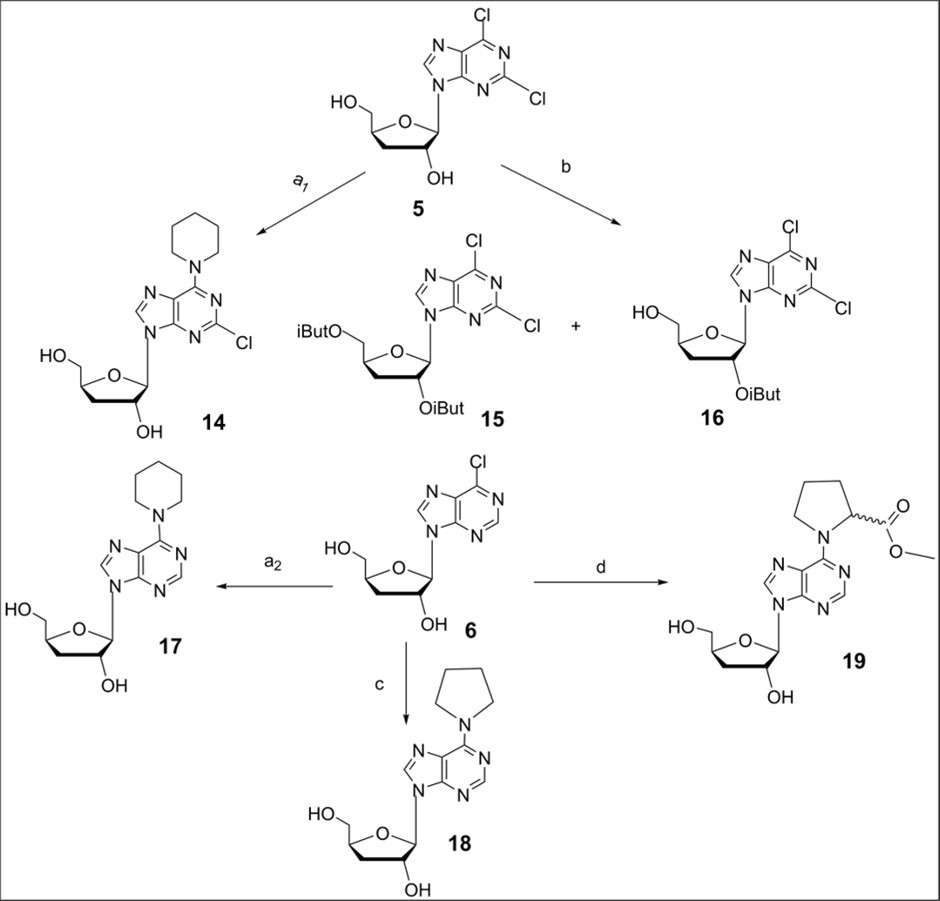
Download figure
Biological evaluation of in vitro antiproliferative activity of purine 3′-deoxyribonucleosides.
A series of purine modified 3′-deoxyribonucleosides prepared by the biocatalytic route and chemical transformations in the heterocyclic base of halogenated nucleosides were tested in vitro on two cancer cell lines, HL-60 (chronic myelogenous leukemia) and K562 (promyelocytic leukemia), compared to cladribine as positive control using MTS assay. The findings of antiproliferative activity for selected purine nucleosides were summarized in Table 2.
Purine 3′-deoxyribonucleosides mono- and disubstituted in the heterobase, acylated in carbohydrate moiety were studied as potential inhibitors of the growth of leukemia cells in cultures. Firstly, antiproliferative activity of dihalogenated and monohalogenated at 2 and 6-positions of purine nucleosides was evaluated in the leukemic cell line HL-60 using the MTS assay 34, 35. Among tested halogenated purine nucleosides, 2,6-chloro (5), 6-chloro (6) and 6-chloro-2-fluoro-purine (7) 3′-deoxyribonucleosides showed ability to inhibit cell growth in HL-60 cells, and 2,6-chloropurine derivative exhibited higher potency (IC50 = 8.9 μM) than nucleosides 6 and 7 (IC50 values of 50μM and 72 μM, respectively). 3′-Deoxy-β-D-ribofuranosyl N6-pyperidin-1-yl-2-chloropurine (14) was inactive in this cell line. 2-Fluorocordycepin displayed activity against the HL-60 cells and 50% of inhibition by this known nucleoside with anticancer and antibacterial activities was observed for concentration of 16.4 μM compared to cladribine (IC50 < 1.0 μM) used as positive control. It should be noted that 3′,5′-di-O-isobutyryl derivative of 2,6-chloropurine 3′-deoxynucleoside 15 and 2′-O-acylated nucleoside 16 demonstrated obvious inhibitory effects withIC50 values of 4.0μM and 2.4 μM towards HL-60 cells in a series of tested purine nucleosides (Table 2). In addition, inhibitory effects of acylated nucleosides of 2,6-chloropurine 15 and 16 were higher than that of the parent nucleoside 5 in HL-60 cells. N6-Monosubstituted purine 3′-deoxyribonucleosides 17, 18 and 19 with pyrrolidine, piperidine or proline substituents, as analogues of cordycepin, were inactive in the cell line HL-60. Three nucleosides 16, 15 and 12 were tested in another leukemic cell line and they showed activity against K562 cells with IC50 values in the range of 19.6 - 49.9 μM in comparison with cladribine (IC50 = 2.5 μM). Purine and pyrimidine 3´-deoxyribonucleosides with natural bases have been reported to exhibit in vitro cytostatic activities 5, 29. In addition, previous biological studies of 5´- and 2´-O-acetylated cytosine 3´-deoxynucleosides and cordycepin derivatives with acetyl groups in the carbohydrate moiety showed that these nucleoside derivatives as potential prodrugs had interesting anticancer activity, cardioprotective or antitumor effects 9, 36. In this context, antiproliferative activity of 2,6-chloropurine 3′-deoxyribonucleoside derivatives 15 and 16 with isobutyryl groups (Table 2) may be consistent with increase of their lipophilicity supporting the high cell penetration. Calculated values of lipophilicity, in silico ADME parameter, were prepared for a set of tested nucleosides (Table 2). In the case of nucleosides 15 and 16, the values of M Log P were 1.97 and 1.07 which is much higher than those for the parent nucleoside 5 (0.09) or purine 3´-deoxyribonucleosides modified in the base. A possible antitumor mechanism of purine modified nucleosides under consideration as well as the 3´-deoxyribonucleosides with natural bases probably includes the formation of nucleoside 5´-O-triphosphates 5, 6, 29 and their antiproliferative activity may correlate with substrate properties of 3´-deoxyribonucleosides for cell kinases in cancer cells.
Table 2. In vitro antiproliferative activities (IC50) of the nucleosides 5-16 against the leukemia cell lines HL-60 and K562| Compound | HL-60 | K-562 | M Log P (TPSA) |
| 5 | 8.9 | ND | 0.09 (93.29) |
| 6 | >50 | ND | -0.46 (93.29) |
| 7 | 72.0 | ND | -0.05 (93.29) |
| 12 | 16.4 | 49.9 | -0.72 (119.31) |
| 14 | - | ND | 0.34 (96.53) |
| 15 | 4.0 | 21.0 | 1.97 (105.43) |
| 16 | 2.4 | 19.6 | 1.07 (99.36) |
| Cladribine | <1.0 | 2.5 | -0.57 (119.31) |
Conclusion
A chemoenzymatic route to novel and the known bioactive purine 3´-deoxyribonucleosides such as cordycepin and its 2-fluoro derivative was explored starting from 3´-deoxyuridine prepared by the nitrite-assisted deamination reaction of 3´-deoxycytidine. It was found that 3´-deoxyuridine can be used as a good donor of the carbohydrate moiety with generation of intermediate 3-deoxy-D-ribofuranose-1-phosphate in the presence of excess of recombinant UP E.coli in the tested enzymatic syntheses of a series of purine 3´-deoxyribonucleosides. Recombinant PNP was shown to possess a broad substrate specificity towards 2,6-disubstituted purines used in transglycosylation reactions. Enzymatic synthesis resulted in good or moderate yields of purine modified nucleosides from the purine bases and 3´-deoxyuridine in the presence of excess of UP and PNPE.coli in potassium phosphate buffer under mild heating. Prepared di- and monohalogenated purine nucleoside derivatives were used as valuable precursors for synthesis of various modified nucleosides. Novel N6-modified cordycepin analogues were obtained from chlorinated purine 3-deoxynucleosides. Antiproliferative activities of a set of purine substituted 3′-deoxyribonucleosides have been evaluated in vitro on two cancer cell lines, HL-60 and K562. Among tested modified purine nucleosides, 2,6-dichlorinated purine 3′-deoxyribonucleosides with 2′,5′- or 2′-O-isobutyryl groups displayed significant antitumor effects in leukemia HL-60 cells. Interestingly, acylated 2,6-chloropurine 3'-deoxynucleosides as well as 2-fluorocordycepin demonstrated inhibitory effects against two leukemic cell lines.
Experimental part
General information
Column chromatography was performed on silica gel 60 H (70-230 mesh; Merck, Darmstadt, Germany), and thin-layer chromatography (TLC) on Merck silica gel aluminum 60 F254 precoated plates. The anhydrous solvents were distilled over CaH2, P2O5 or magnesium prior to the use. All commercially available reagents were used without further purification. 1H, 13C, and 19F NMR spectra were recorded in CDCl3, CD3OD and DMSO-d6 with a Bruker Avance-500-DRX spectrometer at 500.13, 126.76 and 470.59 MHz, respectively. 1H and13C NMR chemical shifts (δ, ppm) are relative to internal chloroform peak (7.26 ppm for 1H and 77.0 for 13C NMR). Chemical shifts are also reported downfield from internal SiMe4 (1H) or external CFCl3 (19F). Splitting patterns were reported as follows: s: singlet, d: doublet, t: triplet, m: multiplet. J values are reported in Hz. Melting points were determined on a Boetius apparatus and were uncorrected. Mass spectra were recorded on HPLC-Accela with LCQ Fleet mass-detector (Thermo electron corporation, USA), using ESI (electrospray ionization). Solutions of recombinant E.coli UP and PNP phosphorylases in 10-15 mM potassium phosphate buffer (pH 7.0) with activities 4300 and 12500 units per ml, respectively, were prepared in laboratory of molecular biotechnology of Institute of Microbiology of National Academy of Sciences of Belarus. Activities of enzymes were evaluated spectrophotometrically by conversions of substrates, inosine and uridine, with E.coli PNP and UP, respectively, to hypoxanthine and uracil in phosphate buffer. A unit of PNP and UPE.coliacivity was defined at the amount of enzyme sufficient to produce 1ϻM of base in 1min under reaction conditions.
Chemoenzymatic synthesis of purine 3′-deoxyribonucleosides.
Synthesis of 3′-deoxyuridine (3) by deamination reaction of 3′-deoxycytidine (1).
a1. 3'-Deoxycytidine (1, 1.5 g, 6.6 mmol) 23, 11 was dissolved in 11 ml glacial acetic acid and then sodium nitrite (2.4 g, 35 mmol) was added to prepared solution. The reaction mixture was stirred at room temperature. After 48 h, sodium nitrite (1.0 g, 14.5 mmol) was added to prepared suspension and then stirring was continued for 48 h at rt. The reaction mixture was diluted with methanol, the prepared solution was filtered off and evaporated, co-evaporated with toluene. The prepared residue was purified by silica gel column chromatography using mixtures of chloroform : methanol from 15:1 to 2:1 to give 1-(5-О-acetyl-3-deoxy-β-D-ribofuranosyl)uracil (2) (0.074 g, 5%) as a colorless oil.1H NMR(500 MHz, CDCl3) δ ppm 7.79 (d,1H, J = 8.2 Hz, H-6), 5.72 (2H, br. d, H-1′ and H-5), 4.72−4.77 (m, 1 H, H-4), 4.49 (d, 1 H, J= 5.0 Hz, H-2), 4.4 (1H, dd, J5′,4′ = 4.4 Hz, J5′,5′ = 12.7 Hz, H-5′), 4.35 (1H, dd, J5′′, 4′ = 2.7 Hz, H-5′′), 2.12 (3H, s, CH3CO), 2.49 (1H, ddd, J = 1.5, 5.1, 13.7 Hz, H-3′), 1.86 (1H, ddd, J = 5.7, 10.2, 13.1 Hz, H-3′′). 13C NMR (126 MHz, CDCl3) δ = 170.4 (CH3CO), 163.6, 151.2, 139.2, 101.9 (C-4, C-2, C-6, C-5), 94.0 (C-1'), 79.4 (C-4'), 76.5 (C-2'), 64.2 (C-5'), 32.8 (C-3'), 29.7 (CH3CO). LC-MS (ESI+): m/z calcd for C11H14N2O6M+Na 293.1, found 293.1.
and 3'-deoxyuridine(3) (0.554 g, 40%) as a white solid. M.p. 177-1790C.
1H NMR (500 MHz, D2O): δ 7.76 (1H, d, J = 8.0 Hz, H-6), 5.67 (1H, d, J = 8.0 Hz, H-5), 5.64 (1H, d, J1′,2′ = 1.3 Hz, H-1′), 4.34-4.39 (2H, m, H-2′ и H-4′), 3.79 (1H, dd, J5′,4′ = 3.8 Hz, J5′,5′ = 12.7 Hz, H-5′), 3.59 (1H, dd, J5′′, 4′ = 4.9 Hz, H-5′′), 1.85-1.88 (2H, m, H-3′ and H-3''). 13C NMR (126 MHz, D2O): δ = 166.0, 151.5, 141.5, 101.4 (C-4, C-2, C-6, C-5), 92.4 (C-1'), 81.6 (C-4'), 75.4 (C-2'), 62.0 (C-5'), 32.6 (C-3'). LC-MS (ESI+): m/z calcd for C9H12N2O4M+Na 235.1, found 235.1.
a2. 3'-Deoxycytidine (1, 0.12 g, 0.56 mmol) was dissolved in 2.4 ml 50% aq. acetic acid and then sodium nitrite (0.164 g, 2.38 mmol) was added to prepared solution. The reaction mixture was stirred at room temperature for 24 h (TLC monitoring CHCl3:MeOH -2:1). The stirring was continued for 30 min at 400C, then pH of the solution was adjusted to pH 7.0 with 25% aq. NH3. The reaction mixture was evaporated under reduced pressure, coevaporated with ethanol, a mixture of ethanol-toluene (1:1). The prepared residue was purified by silica gel column chromatography using mixtures of chloroform: methanol from 10:1 to 2:1 to give 3'-deoxyuridine (3) (0.062 g, 52%) as white solid.
9-(3-deoxy-β-D-ribofuranosyl)-2,6-dichloropurine (5)
2,6-Dichloropurine (0.103 g, 0.609 mmol) and 3′-deoxyuridine (3, 0.278 g, 1.22 mmol) were dissolved in 10 mM K-phosphate buffer (pH 7.4, 10 ml), then uridine phosphorylase (5600 units, 1.3 ml) and purine nucleoside phosphorylase (7000 units, 0.56 ml) were added. The reaction mixture was stirred at 40 0C for 48 h (TLC monitoring CHCl3:MeOH (4:1)), then methanol was added and solvents were removed under reduced pressure. The residue was dissolved in hot methanol, the prepared solution was filtered off and co-evaporated with silica gel and the powdered residue was purified by column chromatography on silica gel using mixtures of chloroform:methanol from 35:1 to 4:1. 9-(3-deoxy-β-D-рибофуранозил)-6-chloro-2-fluoropurine (5) was obtained as a white solid (0.156 g, 84%). M.p. 165-170 0C.
1H NMR (500 MHz, DMSO-d6) δ ppm 8.96 (1H, s, H-8), 5.94 (1H, br.s, H-1′), 5.77 (1H, d, J = 4.0 Hz, OH-2′), 5.10 (1H, t, J = 5.3 Hz, OH-5′), 4.55 (1H, br.d, H-2′), 4.36-4.43 (1H, m, H-4′), 3.73 (1H, dd, J5′,4′= 3.0 Hz, J5′,5′ = 12.2 Hz, H-5′), 3.52 (1H, dd, J5′′, 4′ = 3.6 Hz, H-5′′), 2.16 (1H, ddd, H-3′), 1.85 (1H, ddd, H-3′′). 13C NMR (126 MHz, DMSO-d6) δ = 153.1, 151.4, 150.1, 146.5, 131.5, 91.9, 82.3, 75.3, 62.0, 33.5. LC-MS (ESI+): m/z calcd for C10H10N4O3Cl2M + Na 327.0, found 327.0.
9-(3-deoxy-β-D-ribofuranosyl)-6-chloropurine (6)
6-Chloropurine (0.075 g, 0.485 mmol) and 3′-deoxyuridine (3, 0.22 g, 0.97 mmol) were dissolved in 10 mM K-phosphate buffer (pH 7.4, 7 ml), then uridine phosphorylase (3920 units, 0.91 ml) and purine nucleoside phosphorylase (4900 units, 0.39 ml) were added. The reaction mixture was stirred at 40 0C for 72 h (TLC monitoring CHCl3:MeOH - 4:1), then methanol was added and solvents were removed under reduced pressure. The residue was dissolved in hot methanol, the prepared solution was filtered off and co-evaporated with silica gel and the powdered residue was purified by column chromatography on silica gel using mixtures of chloroform : methanol from 35:1 to 20:1. 9-(3-deoxy-β-D-ribofuranosyl)-6-chloropurine (6) (0.098 g, 73%) was obtained as a white solid. M.p.145-149 0C. 1H NMR (500 MHz, СD3OD): δ ppm 8.95 (1H, s, H-8), 8.75 (1H, s, H-2), 6.15 (1H, d, J1′,2′ = 1.6 Hz, H-1′), 4.73-4.77 (1H, m, H-2′), 4.56-4.60 (1H, m, H-4′), 3.66 (1H, dd, J5′,4′ = 2.7 Hz, J5′,5′ = 12.3 Hz, H-5′), 3.71 (1H, dd, J5′′, 4′ = 3.5 Hz, H-5′′), 2.38 (1H, ddd, H-3′), 2.03 (1H, ddd, H-3′′). 13C NMR (126 MHz, СD3OD): δ = 151.5 (C-2), 145.2 (C-8), 151.0, 149.8, 131.5 (C-6, C-5, C-4), 92.3 (C-1'), 81.9 (C-4'), 75.7 (C-2'), 62.1 (C-5'), 32.8 (C-3'). LC-MS (ESI+): m/z calcd for C10H11N4O3Cl M+Na 293.1, found 293.1.
9-(3-deoxy-β-D-ribofuranosyl)-6-chloro-2-fluoropurine (7)
6-Chloro-2-fluoropurine (0.017 g, 0.1 mmol) and 3'-deoxyuridine (3, 0.023 g, 0.1 mmol) were dissolved in 10 mM K-phosphate buffer (pH 7.4, 1 ml), then uridine phosphorylase (560 units, 0.13 ml) and purine nucleoside phosphorylase (750 units, 0.06 ml) were added. The reaction mixture was stirred at 40 0C for 48 h, (TLC monitoring CHCl3:MeOH - 8:1), then methanol was added and solvents were removed under reduced pressure. The residue was dissolved in hot methanol, the prepared solution was filtered off and co-evaporated with silica gel and the powdered residue was purified by column chromatography on silica gel using mixtures of chloroform: methanol from 30:1 to 15:1. 9-(3-deoxy-β-D-ribofuranosyl)-6-chloro-2-fluoropurine (7) (0.009 g, 32%) was obtained as a white amorphous solid. 1H NMR (500 MHz, DMSO-d6) δ ppm 8.92 (1H, s, H-8), 5.9 (1H, br.s, H-1′), 5.78 (1H, d, J = 4.0 Hz,OH-2′), 5.10 (1H, t, J = 5.3 Hz, OH-5′), 4.54-4.57 (1H, m, H-2′),4.38-4.42 (1H, m, H-4′), 3.74 (1H, ddd, J5′,4′ = 3.3, J5′,5′ = 12.2, J5′,5′OH = 5.1 Hz, H-5′), 3.53 (1H, ddd, J5′′, 4′ = 3.9, J5′,5′OH = 5.4 Hz, H-5′′), 2.18 (1H, ddd, J = 5.2, 8.9, 13.1 Hz, H-3′), 1.85 (1H, ddd, J = 1.5, 5.4, 13.1 Hz, H-3′′). 13C NMR (126 MHz, CD3OD) δ = 156.5 (C-2, d, J = 204.4 Hz), 153.48 (C-6, d, J = 17.3 Hz), 150.7 (C-4, d, J = 18.0 Hz) , 146.4 (C-8), 131.0 (C-5, d, J= 4.5 Hz ), 91.96 (C-1, d, J = 5.4 Hz), 82.34 (C-4, d, J = 2.5 Hz ), 75.39 (C-2, d, J=3.4 Hz), 62.0 (d, C-5, J = 3.7 Hz) , 33.5 (C-3, d, J = 6.3 Hz). 19F NMR (470.59 MHz, DMSO-d6) δ ppm -51.96 (s, F-2). LC-MS (ESI+): m/z calcd for C10H10N4O3ClF M+Na 311.1, found 311.1.
9-(3-deoxy-β-D-ribofuranosyl)-6-chloro-2-aminopurine (8)
6-Chloro-2-aminopurine (0.02 g, 0.118 mmol) and 3′-deoxyuridine (3, 0.053 g, 0.236 mmol) were dissolved in 10 mM K-phosphate buffer (pH 7.4, 2 ml), then uridine phosphorylase (1120 units, 0.26 ml) and purine nucleoside phosphorylase (1400 units, 0.11 ml) were added. The reaction mixture was stirred at 40 0C for 72 h, then methanol was added and solvents were removed under reduced pressure. The residue was dissolved in hot methanol, the prepared solution was filtered off and co-evaporated with silica gel and the powdered residue was purified by column chromatography on silica gel using mixtures of chloroform : methanol from 35:1 to 16:1. 9-(3-deoxy-β-D-ribofuranosyl)-6-chloro-2-aminopurine (8) (0.022 g, 65%) was obtained as a white solid. M.p. 181-185 0C.
1H NMR (500 MHz, DMSO-d6) δ ppm 8.38 (1H, s, H-8), 6.97 (2H, br.s, NH2), 5.78 (1H, d, J1′,2′ = 1.5 Hz, H-1′), 5.64 (1H, d, J = 4.2 Hz,OH-2′), 5.10 (1H, t, J = 5.3 Hz, OH-5′), 4.48-4.65 (1H, m, H-2′), 4.32-4.65 (1H, m, H-4′), 3.67 (1H, dd, J5′,4′ = 3.4 Hz, J5′,5′ = 11.7 Hz, H-5′), 3.51 (1H, dd, J5′′, 4′ = 4.2 Hz, H-5′′), 2.24 (1H, ddd, H-3′), 1.88 (1H, ddd, H-3′′). 13C NMR (126 MHz, DMSO-d6) δ = 160.2, 154.0, 149.9, 141.4, 133.9, 123.9, 90.7, 81.4, 75.2, 62.7, 34.5. LC-MS (ESI+): m/z calcd for C10H12N5O3Cl [M+H] 286.1, found 286.1; M+Na 308.1, found 308.1.
9-(3-deoxy-β-D-ribofuranosyl)-6-thioguanine (9)
6-Thioguanine (0.018 g, 0.11 mmol) and 3′-deoxyuridine (3, 0.049 g, 0.22 mmol) were stirred in 10 mM K-phosphate buffer (pH 7.4, 3.5 ml) under heating 45-50 0C, then to prepared mixture at 40 0C uridine phosphorylase (1670 units, 0.39 ml) and purine nucleoside phosphorylase (2120 units, 0.17 ml) were added. The reaction mixture was stirred for 48 h (TLC monitoring CHCl3:MeOH - 8:1) at 40 0C, then methanol was added and solvents were removed under reduced pressure. The residue was dissolved in hot methanol, the prepared solution was filtered off and co-evaporated with silica gel and the powdered residue was purified by column chromatography on silica gel using mixtures of chloroform : methanol from 25:1 to 5:1. 9-(3-deoxy-β-D-ribofuranosyl)-6-thioguanine (9) (0.009 g, 30%) was obtained as a white amorphous solid.
1H NMR (500 MHz, DMSO-d6) δ ppm 8.10 (1H, s, H-8), 6.80 (2H, br.s, NH2), 5.64 (1H, d, J1′,2′ = 1.9 Hz, H-1′), 5.58 (1H, d, J = 4.2 Hz, OH-2′), 4.9 (1H, t, J = 5.3 Hz, OH-5′), 4.39-4.41 (1H, m, H-2′), 4.25-4.30 (1H, m, H-4′), 3.61 (1H, ddd, J5′,4′ = 3.3 Hz, J5′,5′ = 11.6 Hz, H-5′), 3.46 (1H, ddd, J5′′, 4′ = 3.9 Hz, H-5′′), 2.11 (1H, ddd, J = 13.4, 9.2, 5.7 Hz, H-3′), 1.84 (1H, ddd, J = 2.4, 6.1, 13.4 Hz, H-3′′). 13C NMR (126 MHz, DMSO-d6) δ = 165.8, 153.4, 147.8 (C-6, C-5, C-4), 138.6 (C-8), 128.7 (C-2), 90.4 (C-1′), 81.2 (C-4′), 75.2 (C-2′), 62.7 (C-5′), 34.7 (C-3′). LC-MS (ESI+): m/z calcd for C10H13N5O3S [M+H] 284.1, found 284.2.
3´-Deoxyadenosine (10)
Adenine (0.05 g, 0.37 ммоль) and 3′-deoxyuridine (3, 0.17 g, 0.745 mmol) were stirred in 10 mM K-phosphate buffer (pH 7.4, 3.7 ml) under heating 45-50 0C, then to prepared mixture at 40 0C uridine phosphorylase (2072 units, 0.48 ml) and purine nucleoside phosphorylase (2590 units, 0.21 ml) were added. The reaction mixture was stirred at 40 0C. Progress of the nucleoside formation was monitored by TLC (CH2Cl2:MeOH - 8:1). After 72 h, methanol was added to the reaction mixture and solvents were removed under reduced pressure. The residue was treated with hot methanol, the precipitate was filtered off and washed by methanol. The filtrate was co-evaporated with silica gel and the powdered residue was purified by column chromatography on silica gel using mixtures of ethyl acetate : ethanol from CH2Cl2-MeOH from 35:1 to 6:1 as eluent. 9-(3-deoxy-β-D-ribofuranosyl)-adenine (10) (0.065 g, 70%) was obtained as a white amorphous solid.
1H NMR (500 MHz, СD3OD): δ 8.43 (1H, s, H-8), 8.20 (1H, s, H-2), 5.97 (1H, d, J1′,2′ = 2.7 Hz, H-1′), 4.69-4.72 (1H, m, H-2′), 4.51-4.55 (1H, m, H-4′), 3.93 (1H, dd, J5′,4′ = 2.6 Hz, J5′,5′ = 12.4 Hz, H-5′), 3.66 (1H, dd, J5′′, 4′ = 3.3 Hz, H-5′′), 2.38 (1H, ddd, H-3′), 2.06 (1H, ddd, H-3′′). 13C NMR (126 MHz, СD3OD): δ = 156.0, 152.2, 139.7, 119.2 (C-6, C-5, C-4, C-3), 148.5 (C-8), 92.2 (C-1'), 81.6 (C-4'), 75.2 (C-2'), 62.9 (C-5'), 33.1 (C-3'). LC-MS (ESI+): m/z calcd for C10H13N5O3[M+H] 252.1, found 252.1
9-(3-deoxy-β-D-ribofuranosyl)-2-chloroadenine (11)
2-Chloroadenine (0.04 g, 0.219 mmol) and 3′-deoxyuridine (3, 0.06 g, 0.131 mmol) were stirred in 10 mM K-phosphate buffer (pH 7.4, 3 ml) under heating 50-550C, then to prepared mixture at 40 0C uridine phosphorylase (1120 units, 0.26 ml) and purine nucleoside phosphorylase (1400 units, 0.11 ml) were added. The reaction mixture was stirred at 40 0C. Progress of the nucleoside formation was monitored by TLC (CHCl3:MeOH -8:1). After 72 h, methanol was added to the reaction mixture and solvents were removed under reduced pressure. The residue was treated with hot methanol, the precipitate was filtered off and washed by methanol. The filtrate was evaporated and the residue was chromatographed on silica gel using mixtures of chloroform : methanol from 30:1 to 5:1 as eluent. 9-(3-deoxy-β-D-ribofuranosyl)-2-chloroadenine (11) (0.016 g, 24%) was obtained as a white amorphous solid.
1H NMR (500 MHz, DMSO-d6) δ ppm 8.38 (1H, s, H-8), 7.82 (2H, br s, NH2), 5.80 (1 H, d, J = 2.2 Hz, H-1), 5.69 (1H, d, J = 4.1 Hz, OH-2′), 5.02 (1H, t, J = 5.47 Hz, OH-5′), 4.49−4.55 (1 H, m, H-2), 4.31−4.39 (1H, m, H-4), 3.68 (1H, ddd, J = 12.8, 5.4, 3.2 Hz, H-5′), 3.51 (1H, ddd, J = 11.9, 5.4, 3.9 Hz, H-5′′), 2.21 (1H, ddd, J = 13.20, 8.91, 5.68 Hz, H-3'), 1.90 (1H, ddd, J = 13.07, 6.25, 2.91 Hz, H-3''). 13C NMR (126 MHz, DMSO-d6) δ = 156.6, 152.9, 149.8, 139.3, 117.9, 90.5, 80.9, 74.6, 62.1, 33.7. LC-MS (ESI+): m/z calcd for C10H12N5O3Cl [M+H] 286.1, found 286.1.
9-(3-deoxy-β-D-ribofuranosyl)-2-fluoroadenine (12)
Method A. 2-Fluoroadenine (0.034 g, 0.219 mmol) and 3′-deoxyuridine (3, 0.1 g, 0.438 mmol) were stirred in 10 mM K-phosphate buffer (pH 7.4, 3 ml) under heating 45-50 0C, then to prepared mixture at 40 0C uridine phosphorylase (1680 units, 0.39 ml) and purine nucleoside phosphorylase (2100 units, 0.17 ml) were added. The reaction mixture was stirred at 40 0C for 72 h. Progress of the nucleoside formation was monitored by TLC (ethyl acetate-acetone-water – 7:3:0.5), methanol was added to the reaction mixture and solvents were removed under reduced pressure. The residue was treated with hot methanol, the precipitate was filtered off and washed by methanol. The filtrate was co-evaporated with silica gel and the powdered residue was purified by column chromatography on silica gel using mixtures of ethyl acetate:ethanol from 50:1 to 15:1 as eluent. 9-(3-deoxy-β-D-ribofuranosyl)-2-fluoroadenine (12) (0.023 g, 40%) was obtained as a white solid.
1H NMR (500 MHz, DMSO-d6) δ ppm 8.32 (1H, s, H-8), 5.74 (1H, d, J1′,2′ = 1.8 Hz, H-1′), 5.67 (1H, d, J = 4.0 Hz,OH-2′), 5.02 (1H, t, J = 5.5 Hz, OH-5′), 4.39 (1H, t, H-2′), 4.25-4.34 (1H, m, H-4′), 3.65 (1H, ddd, H-5′), 3.50 (1H, ddd, H-5′′), 2.18 (1H, ddd, H-3′), 1.85(1H, ddd, H-3′′).13C NMR (126 MHz, DMSO-d6): δ = 159.0 (d, J = 203.4 Hz, C2),158.04 (d, J = 20.9 Hz, C6), 150.58 (d, J = 20.2 Hz, C4), 139.72 (C8), 117.83 (C5), 91.14 (C-1′), 81.42 (C-4′), 75.25 (C-2′), 62.78 (C-5′), 34.32 (C-3′). 19F NMR (470.59 MHz, DMSO-d6) δ ppm - 53.2 (s, F-2). LC-MS (ESI+): m/z calcd for C10H12N5O3F [M+H] 270.1, found 270.1; M+ Na. 292.1, found 292.1.
Method B. 2-Fluoroadenosine (13, 0.05 g, 0.175 mmol) 32, 33 and 3′-deoxyuridine (3, 0.073 g, 0.32 mmol) were dissolved in 7.2 ml water and 1 ml 50 mM K-phosphate buffer under stirring and prepared solution of a mixture of nucleosides in 5 mM K-phosphate buffer (pH 7.2) was heated 450-50 0C, then uridine phosphorylase (5600 units, 1.3 ml) and purine nucleoside phosphorylase (7000 units, 0.56 ml) were added at 40 0C. The reaction mixture (the overall volume 10 ml) was stirred at 40 0C for 72 h. Progress of the nucleoside formation was monitored by TLC (ethyl acetate-EtOH-H2O – 7:1:0.5), methanol was added to the reaction mixture and solvents were removed under reduced pressure. The residue was treated with hot methanol, the precipitate was filtered off and washed by methanol. The filtrate was co-evaporated with silica gel and the powdered residue was purified by column chromatography on silica gel using mixtures of ethyl acetate : ethanol from 50:1 to 15:1 as eluent. 9-(3-deoxy-β-D-ribofuranosyl)-2-fluoroadenine (12) (0.033 g) was obtained as a white solid. Yield of the target nucleoside made up 57% according to HPLC analysis for two fractions after column chromatography - 15 mg (91%) and 18 mg (74%) using C18 Column Performance 4.6 x 75 mm, 3.5μ, flow rate 0.5 ml/min, detection at 260 nm, A. 0.1% aq. TFA, B. gradient 10→95% 0.1% aq. TFA in MeCN/H2O.
Synthesis of purine modified 3′-deoxynucleosides from halogenated purine 3′-deoxyribonucleoside 5 and 6.
2-Chloro-6-(pyperidin-1-yl)-9-(3-deoxy-β-D-ribofuranosyl)-purine (14)
To a solution of nucleoside 5 (0.025 g, 0.082 mmol) in absolute ethanol (2 mL), piperidine (0.02 mL, 0.20 mmol) and DIPEA (0.025 ml, 0.14 mmol) were added, and the resulting mixture was stirred for 40 min at room temperature. The reaction mixture was stirred at 85 0C for 2 h, cooled to room temperature then was concentrated in vacuo. The residue was purified by silica gel column chromatography using mixtures of chloroform : methanol from 50:1 to 44:1 to give 14 (0.028 g, 98%) as a white solid. M.p. 91-95 0C. 1H NMR (500 MHz, DMSO-d6) δ ppm 8.4 (1 H, s, H-8), 5.8 (1H, d, J1′,2′ = 1.7 Hz, H-1′), 5.68 (1H, d, OH-2′), 5.02 (1H, t, OH-5′), 4.45 (1H, br.m, H-2′), 4.31-4.36 (1H, m, H-4′), 3.60 (1H, ddd, H-5′), 3.49 (1H, ddd, H-5′′), 3.29 [(4H, br.s, --N-(CH2-)2], 2.14 (1H, ddd, J = 5.5, 8.0, 13.1 Hz, H-3′), 1.85 (1H, ddd, = 1.9, 4.5, 13.1 Hz, H-3′′), 1.62-1.66 (2H, m, CH2-), 1.51-1.58 (4H, m, 2x CH2-).13C NMR (126 MHz, DMSO-d6) 153.6, 153.1, 151.4, 138.5, 118.6, 91.0, 81.6, 75.4, 62.6, 39.5, 26.1, 24.5. LC-MS (ESI+): m/z calcd for C15H20N5O3Cl [M+H] 354.1, found 354.2; M+Na 376.1, found 376.2.
2,5-Di-О-isobutyroyl-9-(3-deoxy-β-D-ribofuranosyl)-2,6-dichloropurine (15) and 2-О isobutyroyl-9-(3-deoxy-β-D-ribofuranosyl)-2,6-dichloropurine (16)
Isobutyric anhydride (0.066 mL, 0.36 mmol) and DIPEA (0.013 ml, 0.09 mmol) were added to a solution of nucleoside 5 (0.06 g, 0.198 mmol) in anhydrous acetonitrile (4.5 mL), and the resulting solution was stirred for 20 min at 0 0C. The reaction mixture was stirred for 20 h at room temperature, then was concentrated in vacuo. The residue was purified by silica gel column chromatography using mixtures of chloroform : methanol from 50:1 to 24:1 to give nucleoside 2,5-di-О-isobutyroyl-9-(3-deoxy-β-D-ribofuranosyl)-2,6-dichloropurine(15)(0.009 g, 15%) as oil.
1H NMR (500 MHz, CDCl3) δ ppm 8.3 (1H, s, H-8), 5.94 (1H, d, J = 1.3 Hz, H-1′), 5.60 (1H, br.d, H-2′), 4.64-4.69 (1H, m, H-4′),4.44 (1H, dd, J5′,4′ = 2.7 Hz, J5′,5′ = 12.2 Hz, H-5′), 4.32 (1H, dd, J5′′, 4′ = 5.3 Hz, H-5′′), 2.62-2.66 (2H, m, 2x-CH(CH3)2, 2.56-2.62 (1H, m, H-3′), 2.26 (1H, ddd, J = 14.0, 5.7, 1.4 Hz, H-3′′), 1.2 (6H, dd, 2x-OCH(CH3)2, 1.15 (6H, dd, 2x-OCH(CH3)2. 13C NMR (126 MHz, CDCl3) δ =176.8 and 176.4 [2x (CH3)2CHCO], 153.2, 152.2, 152.1, 144.4, 131.5 (C-6, C-2, C-4, C-8, C-5), 90.5 (C-1'), 79.2 (C-4'), 77.7 (C-2'), 64.2 (C-5'), 33.9 and 33.8 [2 x-COCH(CH3)2], 32.7 (C-3'), 19.1, 18.98, 18.86, 18.79 [2x (CH3)2CHCO]. LC-MS (ESI+): m/z calcd for C18H22N4O5Cl2[M+H] 445.2, found 445.2; M+Na 467.2, found 467.2. and 2-О-isobutyroyl-9-(3-deoxy-β-D-ribofuranosyl)-2,6-dichloropurine (16) (0.018 g, 36%) as oil.
1H NMR (500 MHz, CDCl3) δ ppm 8.37 (1H, s, H-8), 6.0 (1H, d, J = 2.6 Hz, H-1′), 5.50 (1H, dt, H-2′), 4.59 (1H, m, 5′-OH), 4.12 (1H, br.d, H-5′), 3.74-3.80 (1H, m, H-4′), 3.41 (1H, dd, H-5′′), 2.84 (1H, ddd, H-3′), 2.59-2.65 (1H, m, CH(CH3)2, 2.21 (1H, ddd, H-3′′), 1.19 [(3H, d, COCH(CH3)2], 1.18 [(3H, d, COCH(CH3)2]. 13C NMR (126 MHz, CDCl3) δ =176.6 [(CH3)2CHCO], 153.0, 152.4, 152.1, 145.0, 131.7 (C-6, C-2, C-4, C-8, C-5), 91.2 (C-1′), 81.9 (C-4′), 78.1 (C-2′), 62.9 (C-5′), 33.8 COCH(CH3)2, 31.2 (C-3′), 18.85 and 18.81 [(CH3)2CHCO]. LC-MS (ESI+): m/z calcd for C14H16N4O4Cl2M+Na 397.0, found 397.0. and the starting nucleoside (0.018 g, recovery 30% of 5).
6-Pyperidin-1-yl-9-(3-deoxy-β-D-ribofuranosyl)-purine (17)
To a solution of nucleoside 6 (0.025 g, 0.074 mmol) in absolute ethanol (2 mL), piperidine (0.018 mL, 0.185 mmol) and DIPEA (0.023 ml, 0.132 mmol) were added, and the resulting mixture was stirred for 40 min at room temperature. The reaction mixture was stirred at 85 0C for 2 h, cooled to room temperature then was concentrated in vacuo. The residue was purified by silica gel column chromatography using mixtures of chloroform: methanol from 50:1 to 36:1 to give 17 (0.023 g, 98%) as a white solid. M.p. 181-184 0C. 1H NMR (500 MHz, CD3OD) δ ppm 8.27 (1H, s, H-8), 8.14 (1H, s, H-2), 5.88 (1H, d, J1′,2′ = 2.6 Hz, H-1′), 4.62-4.64 (1H, m, H-2′), 4.46-4.50 (1H, m, H-4′), 3.89 (br. s, 4H, -N-CH2), 3.90 (1H, dd, J5′,4′ = 2.5 Hz, J5′,5′ = 12.4 Hz, H-5′), 3.62 (1H, dd, J5′′, 4′ = 3.1 Hz, H-5′′), 2.31 (1H, ddd, J = 6.4, 8.0, 13.1 Hz, H-3′), 2.01 (1H, ddd, J = 3.8, 6.6, 13.1 Hz, H-3′′), 1.69 - 1.74 (2H, m), 1.59 - 1.63 (4H, m). 13C NMR (126 MHz, CD3OD) 153.5, 151.5, 149.3, 137.6, 119.8, 92.1, 81.0, 75.1, 62.7, 33.0, 25.8, 24.4. LC-MS (ESI+): m/z calcd for C15H21N5O3[M+H] 320.3, found 320.3; M+Na 342.1, found 342.1.
6-Pyrrolidin-1-yl-9-(3-deoxy-β-D-ribofuranosyl)-purine (18)
To a solution of nucleoside 6 (0.02 g, 0.074 mmol) in absolute ethanol (2 mL), pyrrolidine (0.012 mL, 0.148 mmol) and triethyl amine (0.023 ml, 0.163 mmol) were added, and the resulting mixture was stirred for 40 min at room temperature. The reaction mixture was stirred at 85 0C for 3 h, cooled to room temperature then was concentrated in vacuo. The residue was purified by silica gel column chromatography using mixtures of chloroform : methanol from 45:1 to 20:1 to give nucleoside 18 (0.022 g, 98%) as a white solid. M.p. 183-186 0C.
1H NMR (500 MHz, CDCl3) δ ppm 8.05 (1H, s, H-8), 7.74 (1H, s, H-2), 6.1 (1H, br.s, OH-2′), 5.57 (1H, d, J1′,2′ = 6.1 Hz, H-1′), 5.18 (1H, br.s, OH-5′), 5.09 (1H, q, H-4′), 4.47 (1H, dd, J = 1.4, J = 9.0 Hz, H-2′), 3.98-4.10 (2H, m,-N-CH2), 3.92 (1H, dd, J5′, 4′ = 1.2 Hz, J5′,5′ = 12.7 Hz, H-5′), 3.54-3.63 (4H, m, H-5′′,-N-CH2), 2.53 (1H, ddd, J = 3.1, 7.5, 12.5 Hz, H-3′), 2.26 (1H, ddd, J = 8.6, 9.0, 12.5 Hz, H-3′′), 2.01-2.09 (2H, m, CH2- ), 1.95-2.00 (2H, m, CH2- ). 13C NMR (126 MHz, CDCl3) 152.6, 151.9, 149.3, 148.2, 138.6, 121.3, 93.2, 80.2, 72.5, 65.1, 48.9, 47.5, 33.7, 29.7, 26.2, 24.3. LC-MS (ESI+): m/z calcd for C14H19N5O3[M+H] 306.1, found 306.1; M + Na 328.1, found 328.1.
6-D/L-proline methyl ether-9-(3-deoxy-β-D-ribofuranosyl)-purine (19)
To a solution of nucleoside 6 (0.03 g, 0.11 mmol) in anhydrous acetonitrile (3 mL), D/L-proline methyl ether hydrochloride (0.02 g, 0.122 mmol) and DIPEA (0.02 ml, 0.11 mmol) were added at 0 0C, and the resulting mixture was stirred for 48 h at room temperature. Then D/L proline methyl ether hydrochloride (0.013 g, 0.167 mmol) and DIPEA (0.029 ml, 0.17 ммоль) were added. After stirring for 48 h the resulting mixture was concentrated in vacuo. The residue was purified by silica gel column chromatography using mixtures of chloroform: methanol from 40:1 to 32:1 to give a mixture of isomeric nucleosides 19 (0.035g, 92%) as oil.
1H NMR (500 MHz, CD3OD, a mixture of diastereomers with D- and L-proline methyl ether at C6 of purine) δ ppm 8.37 (1H, s, H-8), 8.13 (1H, s, H-2), 8.22 (0.91H, s, H-8), 8.17 (0.91H, s, H-2), 5.91 (1H, br.s, H-1′), 5.87 (0.91H, br.s, H-1′), 4.75 (br.s 0.91H, H-2′), 4.62-4.64 (1.97H, m, H-2′ and H-4′), 4.47 (1H, m, H-4′), 4.1-4.25 (2H, 2x- CHCO(OCH3), 3.82- 3.92 (2H, m, 2H-5′), 3.59-3.70 (8H, 2H-5′and 2xOCH3), 2.23-2.44 (5H, H-3′ and proline CH2), 1.85-2.1 (8H, H-3′′ and proline CH2). 13C NMR (127.76 MHz, CD3OD) 153.5, 152.5, 151.6, 151.55, 149.96, 148.77, 138.9, 138.7 (6-proline methyl ether of purine), 92.1 (C-1′), 81.2 and 80.9 (C-2′), 75.2 and 74.9 (C-4′), 62.9 and 62.7 (C-5′), 61.2 and 60.2 (OCH3), 51.57, 51.37 (CH), 49.1, 47.7 (CH2) 33.2 and 32.9 (C-3′), 30.7, 28.7, 24.4, 21.9 (CH2). LC-MS (ESI+): m/z calcd for C16H22N5O5M+ 364.2, found 364.3.
Biological assays of antiproliferative activity
Human cancer cell lines, HL-60 (promyelocytic leukemia) and K-562 (chronic myelogenous leukemia), were obtained from the Institute of Cytology, Russian Academy of Sciences. Cells were maintained in RPMI-1640 supplemented with 10 % fetal bovine serum (HyClone), 100 U/mL penicillin, 100 μg/mL streptomycin, 25 μg/mL amphotericin B. Cultures were incubated at 37 0C in a humidified atmosphere containing 5 % CO₂ in the NU-5840E cell incubator (NuAire). The cells were plated in 96-well plates at a density of 10 × 103 cells per well in 90 μL of medium and allowed to adhere for 1 h. Test compounds were initially prepared as 20 mM stock solutions in DMSO and subsequently diluted in culture medium to the required working concentrations. Cells were then incubated for an additional 72 h. Cellular sensitivities to nucleoside derivatives were measured using the CellTiter 96® AQueous One Solution Cell Proliferation Assay (MTS, Promega Corporation). Following incubation with the MTS reagent for 240 min at 37 0C, absorbance was measured at 492 nm using the Awareness Microplate Reader Stat Fax 3200. The mean absorbance from triplicate wells was calculated, and cell viability was expressed as a percentage of the untreated control using the formula: Viability (%) = (OD_sample / OD_control) × 100, where OD_sample is the absorbance of wells containing the test compound, and OD_control corresponds to wells treated with 0.5 % DMSO (vehicle control). Each experiment was conducted independently three times. IC50 values were calculated using GraphPad Prism software and the results are summarized in Table 2.
References
- 1.Kumar A, S I Khan, Manglani A, Z K Khan, S B Katti. (1994) Synthesis and antifungal activity of 3'-deoxyribonucleosides. , Doi: https://doi.org/10.1080/15257779408011878, Nucleosides & Nucleotides 13, 1049-1058.
- 2.He R, Zhou W. (2024) Application and research progress of cordycepin in the treatment of tumors,Mol. Med. Reports 30, 161-10.
- 3.Hulpia F, K Van Hecke, F da Silva C, G Batista J D da, Maes L. (2018) Discovery of Novel 7-aryl 7-deazapurine 3´-deoxy-ribofuranosyl nucleosides with potent activity against Trypanosoma cruzi, J. Doi: https://doi.org/ 10.1021/acs.jmedchem.8b00999 , Med. Chem. 61 9287-9300.
- 4.V N Barai, A I Zinchenko, L A Eroshevskaya, E V Zhernosek, E De Clerq. (2002) Chemo-Enzymatic Synthesis of 3-Deoxy-β-D-ribofuranosyl Purines, Helv. Chim. Acta, 85 7, 1893-1900.
- 5.Yu G, Peng J, Li L, Yu W, He B. (2024) The role and mechanisms of cordycepin in inhibiting cancer cells, Braz. J. Med. Biol. Res. 57: 13889, 10-1590.
- 6.J B Lee, Radhi M, Cipolla E, R D Gandhi, Sarmad S. (2019) A novel nucleoside rescue metabolic pathway may be responsible for therapeutic effect of orally administered cordycepin, Scientific Reports. 9. 15760-10.
- 7.S K Vodnala, Lundback T, Yeheskieli E, Sjoberg B, Gustavsson A-L. (2013) Structure−Activity Relationships of Synthetic Cordycepin Analogues as Experimental Therapeutics for African Trypanosomiasis, J. Med. Chem. 56, 861−9873. Doi: https://doi.org/10.1021/jm401530a
- 8.A M Rabie. (2022) . Potent Inhibitory Activities of the Adenosine Analogue Cordycepin on SARS-CoV-2 Replication, ACS Omega.7,2960−2969.https://doi.org/10.1021/acsomega.1c05998 .
- 9. (1991) Synthesis and Anticancer Activity of various 3´-deoxy pyrimidine nucleosides analogues and crystal structure of 1-(3-deoxy-β-D-threo-pentofuranosyl)cytosine. J. Med. Chem 34, 693-701.
- 10.Huang S, Liu H, Sun Y, Chen J, Li X. (2018) An effective and convenient synthesis of cordycepin from adenosine. Chem Pap 72, 149-160.
- 11.Marcuccio S, B C Elmes, Holan G, Middleton E J. (1992) Modified Nucleosides. , Doi: https://doi.org/10.1080/0732831920808017816, II. Economical Synthesis of 2′,3′-Dideoxycytidine. Nucleosides & Nucleotides 11, 1695-1701.
- 12.S Y Rhie, Pfleiderer W. (1994) . Nucleosides. LVI. Synthesis and Chemical Modifications of 3’-deoxy pyrimidine nucleosides. Nucleosides & Nucleotides. 13 , Doi: https://doi.org/10.1080/15257779408012162 6, 1425-1452.
- 13.Misra S, Jain S, Avasthi K, D S Bhakun. (1990) . Studies on nucleosides: PART XXVIII. Synthesis of 4-amino (or hydroxy)-6-methylthio-1-(3'-deoxy-β-D-ribofuranosyl)-1-H-pyrazolo[3,4-d] pyrimidines. Nucleosides & Nucleotides , Doi: https://doi.org/10.1080/15257779008043149 9, 837-846.
- 14.Kaspar F, Stone M R L, Neubauer P, Kurreck A. (2021) Route efficiency assessment and review of the synthesis of β-nucleosides: Via N -glycosylation of nucleobases, Green. Doi: https://doi.org/10.1039/DOGC02665D , Chem 23, 37-50.
- 15.Zhou X, Szeker K, Jiao L-Y, Oestreich M, I A Mikhailopulo et al. (2015) Synthesis of 2,6-Dihalogenated Purine Nucleosides by Thermostable Nucleoside Phosphorylases. Doi: https://dx.doi.org/10.1002/adsc.201400966 , Adv Synth. Catal 357, 1237-1244.
- 16.Zhou X, I A Mikhailopulo, Bournazou M N C, Neubauer P. (2015) Immobilization of thermostable nucleoside phosphorylases on MagReSyn®epoxide microspheres and their application for the synthesis of 2,6-dihalogenated purine nucleosides. , Doi: https://doi.org/10.1016/j.molcatb.2015.02.009, J.Mol.Cat.B:Enzym 115, 119-127.
- 17.Cosgrove S, G J Mille. (2022) Advances in biocatalytic and chemoenzymatic synthesis of nucleoside analogues. , Doi: https://doi.org/10.1080/17460441.2022.2039620, Expert Opinion on Drug Discovery 17, 355-364.
- 18.A O Denisova, Y A Tokunova, I V Fateeva, A, V N Leonov. (2017) The Chemoenzymatic Synthesis of 2-Chloro- and 2-Fluorocordycepins, Synthesis. 49. 4853-4860.
- 19.A O, K V Antonov, E A Zorina, M A Simonova, A S Paramonov. (2025) 2-Fluorocordycepin:Chemoenzymatic synthesis and study of anticancer activities in vitro. Rus. , Doi: https://dx.doi.org/10.1011341/Ы1068162025601144, J.Bioorg. Chem 51, 1189-1205.
- 20.A I Beresnev, S V Kvach, G, A I Zinchenko. (2013) Synthesis of 3′-α-fluoronucleosides with using pyrimidine nucleosidephosphorylase Thermus thermophilus and puirine nucleoside. phosphorylase Escherichia coli.,Proceedings of the National Academy of Sciences of Belarus.ser 3, 73-77.
- 21.A I Beresnev, S V Kvach, G, T S Bozhok, E N Kalinichenko et al. (2015) Use of fermentative transglycosylation for preparation of fluoro. nucleosides Proceedings of the National Academy of Sciences of Belarus.ser 2, 66-73.
- 22.D V Burko, L A Eroshevskaya, S V Kvach, A V Shakhbazau, N A Kartel et al. (2010) Chapter 1.Application of recombinant enzymes for the synthesis of pharmaceutically valuable nucleosides and nucleotides/Biotechnology. in Medicine, Foodstuffs, Bicataysis, Environment and Biotechnology// Eds: S.D. Varfolomeev, G.E. Zaikov, L.P. Krylova. – NY: , Nova Science Publishers, Inc.- 1-13.
- 23.G, T V Klennitskaya, E V Zhernosek, I A Mikhailopulo. (2002) Synthesis of Peracylated Derivatives of L-Ribofuranose from D-Ribose and Their Use for the preparation of β-L-Ribonucleosides. Synthesis 253-259.
- 24.J K Novak, Ṧorm F. (1973) Nucleic acid components and their analogues. CLVII. 5'-triphosphates of 3'-deoxyribonucleosides. Coll. Czech. Commun , Doi: https://doi.org/10.1135/CCCC19731173 38, 1173-1178.
- 25.Miah A, C B Reese, Song Q, Sturdy Z, Neidle S. (1998) 2',3'-Anhydrouridine. A useful synthetic intermediate. J. Chem. Soc., Perkin Trans. I.3277–3283. https://doi.org/10.1039/A803563F
- 26.Shapiro R, Yamaguchi H. (1972) Nucleic acid reactivity and conformation deamination of cytosine by nitrous acid, Biochim. Biophys. Acta 281, 50-506.
- 27.Saneyoshi M, Tohyama J, Nakayama C. (1982) Synthesis of Nucleosides and Nucleotides. XIX.1) Synthesis of 3‘-Deoxycytidine 5’-Triphosphate and Ralated 3’-Deoxyribonucleotides from Cordycepin, Chem. Pharm. Bull , Doi: https://doi.org/10.1248/cpb30.2223 30, 2223-2227.
- 28.Shen W, Wang J. (2024) Deamination- or N-Nitrosation-Based Methods for m6A Profiling. , Doi: https://doi.org/10.1002/ijch.202300180, Isr J. Chem 64, 202300180.
- 29.Saneyoshi M, Kohsaka-Ichikawa M, Yahata A, Kimura S, Izuta S. (1995) Synthetic Nucleosides and Nucleotides. XXXV.1)Synthesis and Biological Evaluations of 5-Fluoropyrimidine Nucleosides and Nucleotides of 3-Deoxy-β-D-ribofuranose and Related Compounds, Cherm. Pharm. Bull 43, 2005-2009.
- 30.S V Kvach, L A Eroshevskaya, A I Zinchenko, A V Shakhbazov, N A Kartel’. (2012) Bacterial strain of Escherichia coli producing uridine phosphorylase. Institute of Microbiology of National Academy of Sciences of Belarus, Institute of Genetics and Cytology of National Academy of Sciences of Belarus , BY, In. Cl 7, 12-1.
- 31.S V Kvach, L A Eroshevskaya, A I Zinchenko, A V Shakhbazov, N A Kartel’. (2010) Bacterial strain of Escherichia coli producing purine nucleoside phosphorylase. Institute of Microbiology of National Academy of Sciences of Belarus, Institute of Genetics and Cytology of National Academy of Sciences of Belarus , BY, In. Cl 7, 12-1.
- 32.Krollikiewicz K, Vorbruggen H. (1994) The synhesis of 2-fluoropurine nucleosides, Nucleosides. , Doi: https://doi.org/10.1080/152577794080113271, Nucleotides 13, 673-678.
- 33.V B Berzin, E V Dorofeeva, V N Leonov, A I Miroshnikov. (2009) The preparative method for 2-fluoroadenosine synthesis. Russ J.Bioorg. Chem 35, 193-196.
- 34.A H Cory, T C Owen, J A Barltrop, J G Cory. (1991) Use of an aqueous soluble tetrazolium/formazan assay for cell growth assays in culture. Doi: https://doi.org/10.3727/095535491820873191 , Cancer Commun 3, 207-212.