
Severe Infantile Blount’s Disease in Kumasi, Ghana: A Case Report
Abstract
Blount’s disease, also known as tibia vara, is a developmental disorder involving the posteromedial proximal tibial physis resulting in progressive varus, procurvatum and internal torsion of the affected tibia 1. The condition was first published by Blount in 1973 2. The aetiology of this disease is unkown. However, associations exist between blount’s disease and the Afro-Caribbean race, early age of walking and obesity 3, 4. Furthermore, genetic predisposition has been postulated as well as mechanical loading of the physis 4, 5, 6. Affected children are usually overweight and start walking early. It is bilateral in 80% of cases 7.
Author Contributions
Academic Editor: Shuai Li, Department of Engineering University of Cambridge UK.
Checked for plagiarism: Yes
Review by: Single-blind
Copyright © 2019 Dominic Konadu-Yeboah, et al.
 This is an open-access article distributed under the terms of the Creative Commons Attribution License, which permits unrestricted use, distribution, and reproduction in any medium, provided the original author and source are credited.
Competing interests
The authors have declared that no competing interests exist.
Citation:
Introduction
Blount’s disease, also known as tibia vara, is a developmental disorder involving the posteromedial proximal tibial physis resulting in progressive varus, procurvatum and internal torsion of the affected tibia 1. The condition was first published by Blount in 1973 2. The aetiology of this disease is unkown. However, associations exist between blount’s disease and the Afro-Caribbean race, early age of walking and obesity 3, 4. Furthermore, genetic predisposition has been postulated as well as mechanical loading of the physis. 4, 5, 6 Affected children are usually overweight and start walking early. The role of weight bearing in the pathogenesis of tibia vara was demonstrated in a study by Cook and others 5 who concluded that weight bearing was necessary for the disease to occur, since it is almost never diagnosed before 2 years of age. The disease often manifests after at least 1 year of walking and is never encountered in non-ambulatory children. Blount’s disease is a progressive disorder which if left untreated leads to premature physeal arrest at the proximal posteromedial tibia with a resultant varus deformity of the proximal tibia.
16. It is bilateral in 80% of cases 7 and a cause of significant knee pain and activity limitation among affected children and adolescents.
Pathology
Tibia vara is a disease that involves the epiphysis, physis and the metaphysis 3, 8, 9. Disruption of the normal enchondral ossification in the posteromedial aspect of the proximal metaphysis occurs with defective growth of the posteromedial physis 10, 11. The disruption of growth at the proximal medial tibial physis occurs due to abnormal stresses on the epiphysis in response to the Heuter-Volkman law which states that increased pressure on an epiphysis inhibits growth 5. This concept is reinforced by Delpech’s law which says stimulation of epiphyseal growth occurs by release of pressure 5. It therefore follows that exertion of compressive stress on the medial epiphysis should retard growth whereas reducing stress on the lateral epiphysis should stimulate growth culminating in a varus deformity of the proximal tibia. There are two types of Blount’s disease, infantile form which appears before age 3 years and adolescent type which occurs in children older than 10 years 10.
The late stages of the infantile disease may be accompanied by depression of the medial tibial plateau in addition to the varus and the internal torsion of the tibia 11.
Radiographic Assessment
Weight-bearing radiographs are taken in the anteroposterior and lateral projections of the lower limbs in a standardized form.The disease can be classified using Langenskiold classification system, Figure 1, 12.
Figure 1.Langenskiold staging describes six radiographic stages of the disease based on the degree of epiphyseal depression and metaphyseal fragmentation of the proximal medial tibial epiphysis 13.
Figure2 depicts the measurement of the metaphyseo-diaphyseal angle which is another method for classifying the disease. A line is drawn perpendicular to the long axis of the tibia and another across the metaphyseal flare. The acute angle formed by these two lines should normally not exceed 11 degrees 7.
Figure 2.These classification systems are employed to aid diagnosis, monitor progression and to guide treatment of the disease 13
The metaphyseo-diaphyseal angle (MDA) better correlates with treatment outcomes as compared with the Langenskiold classification system 14.
These classification systems are employed to aid diagnosis, monitor progression and to guide treatment of the disease 13
There have been few reports of successful managament of the late stage of this condition from Africa.
We report our experience in the management of a case of infantile blount’s disease in a boy who was treated operatively at the age of 13 years with the application of the “double corrective osteotomy” surgical technique.
Case Report
A 13-year old boy presented to our hospital with a 10-year history of progressive severe lateral bowing of both legs which was noticed by the mother when the child was 3 years of age. Lateral bowing of the legs had become so severe that his walking had become limited to a few meters, beyond which he developed knee pain. He was borne to a 38-year old multiparous petty trader.
Physical examination revealed a boy who looked well with bilateral tibia vara, procurvatum and internal tibial torsion, Figure 3. There was a palpable posteromedial metaphyseal beak. There were no abnormalities detected in other systems.
Figure 3.Pre-operative photograph of the patient, standing I, and lying, II.
There was a varus angle of 70 degrees as measured by a goniometer when weight-bearing. The range of motion in both knees was 0-100 degrees. X-ray confirmed a Langenskiold stage VI tibia vara on either side, Figure 4.
Figure 4.Fig showing pro-operative radiograph of the left tibia and fibula
He was then prepared for operative correction of the deformity and surgery was performed.
Surgical Technique
Under general anaesthesia with the use of tourniquet, via lateral approach to the fibula, about 3-cm of the proximal one-third of the shaft of the fibular, about 10cm from the head, was excised.
A lateral based closing wedge tibial osteotomy was performed about 5cm distal to the knee joint line, below the tibial tuberosity. A bony wedge of the tibia was removed to correct the varus and the internal torsion of the tibia, overcorrection of 10 degrees of valgus was done to minimize the risk of recurrence. The osteotomy was fixed with an 8-plate using 4.0mm cannulated screws, one in each fragment.
The second osteotomy was performed just below the “beak” under image intensifier control, using a chisel, targeting the intercondylar area of the femur without crossing the articular cartilage, along two parallel guide wires. Care was taken to make the cut parallel to the physis. The knee was placed in flexion during the osteotomy to minimize neurovascular damage.
The bony bridge at the posteromedial physis was totally removed and the resulting defect filled with subcutaneous fat. The medial tibial plateau was then elevated. The wedge of bone removed from the closing wedge tibial osteotomy and the excised fibula were used as a strut to plug the void that resulted from the medial plateau elevation and to support the elevated plateau. The interposed bony material was stabilized using transfixing kirchner wires that were in turn coupled to the circular frame.
The procurvatum and the internal torsion of the tibia were corrected and taylor spatial frame applied to further maintain the correction. The surgical wounds were closed in layers and dressed.
The procedure was done first on the right tibia and fibula and then repeated on the left at the same sitting. The patient was discharged home on post-operative day 10 to mobilise full weight-bearing with a pair of axillary crutches. The patient commenced range of motion exercises of both knee and ankle joints on day 1 post operatively. The taylor spatial frame was removed six months after surgery.
Discussion
The diagnosis of infantile blount’s disease is made clinically and confirmed with plain radiographs of the affected limbs.
The efficacy of bracing as a method of treatment is yet to be proven 4. There are limited reports in the last decade on the use of proximal tibial osteotomy in the correction of this deformity. Acute or gradual correction are both appropriate treatment options 4.
Early surgical intervention at the early stages of the disease results in a low incidence of recurrence of the deformity and reduced knee symptoms at skeletal maturity 15. Patients with blount’s disease should be managed operatively as soon as the disease is diagnosed since the deformities worsen if left untreated 15.
When neglected, blount’s disease is characterized by multiple deformities and a detailed patient-specific treatment plan is mandatory 16. Tibial valgus osteotomy does not offer adequate correction of the deformity and additional elevation of the depressed tibial plateau is essential 16 i. e. ‘double corrective osteotomy’ of the tibia, with medial plateau elevation. It is a demanding and a relatively high-risk procedure. However, in spite of the technical difficulties, it offers favourable results in Langenskiold and Riska stage V or greater 16.
The use of the Taylor Spatial Frame or the Ilizarov Frame are modern treatment mothods that allow gradual correction as well as weight-bearing throughout the treatment period 16. Their main demerits are their relatively high cost and the complexity of their application.
Operative treatment of blount’s disease requires corrective osteotomy, proximal tibila physeal distraction, physeal bar resection and elevation of the medial tibial plateau; lateral hemiepiphyseodesis is recommended particularly in young patiennts 17.
Osteotomy and gradual deformity correction of either infantile or adolescent blount’s disease with the use of a circular external fixation offers satisfactory correction without significant complications 18.
The risk of recurrence of the deformity is related to the poor growth potential of the posteromedial proximal tibial physis 19. Young age of patient and disease severity are the important determinants of deformity recurrence, being 50% for Langenskiold stage III and 69.6% for Langenskiold stage IV respectively. Age 5 seems to be a critical transition in the risk of recurrence of deformity after osteotomy of the tibia.
After high tibial osteotomy or in combination with epiphysiodesis, severe vertical sloping of the metaphyseal defect carries a poor prognosis for success 14. The complications reported in the literature after high tibial osteotomies are: common peroneal nerve palsy, compartment syndrome, haematoma, deep vein thrombosis, osteomyelitis, delayed union, malunion, non-union and infarction of the proximal tibia 20, 21
In 1964, Langenskiold and Riska described elevating the medial plateau in late cases of infantile Blount’s disease 2. Gradual correction using the Ilizarov method for Langenskiold types IV, V and VI has also been described 1. There are various treatment methods of this condition with varying degrees of efficacy and ease of application. Studying the various methods for the correction of neglected blount’s disease will enhance the development of treatment algorithms 22, 23, 24, 25.
In this report we present our correction procedure for Langenskiold Type VI with a single-stage double tibial osteotomy and maintenance of correction of deformity with a Taylor Spatial Frame. Figure 5, Figure 6.
Figure 5.Immediate post-operative picture of the patient lying on the operating table.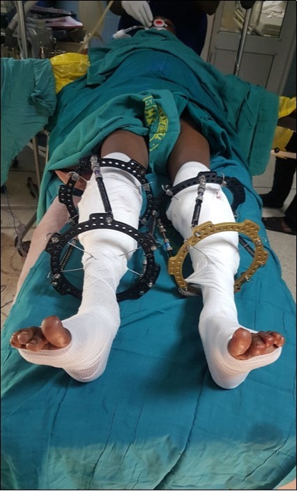
Figure 6.Post-operative x-ray showing the proximal tibia and the fibula osteotomy as well as the Taylor Spatial Frame.
Post-Operative Rehabilitation Regimen
The patient was mobilised full weight-bearing from post-operative day 1 and he commenced quadriceps and hamstrings strengthening exercises as well as active range of motion exercises of both knees on the same day. He was discharged home on post-operative day 10.
The patient developed a right foot drop which resolved after six weeks and a pin tract infection which resolved after 14 days of clindamycin therapy.
There was satisfactory correction of his deformities at his last out-patient visit, 2 years post operatively, with acceptable mobility of the knees, 0- 120 degrees of passive knee motion. Figure 7.
Figure 7.A photograph of the patient, 2 years post-operatively, sitting on a couch with knees flexed, I, and standing on the floor, II, showing correction of varus and torsional deformities of the tibiae.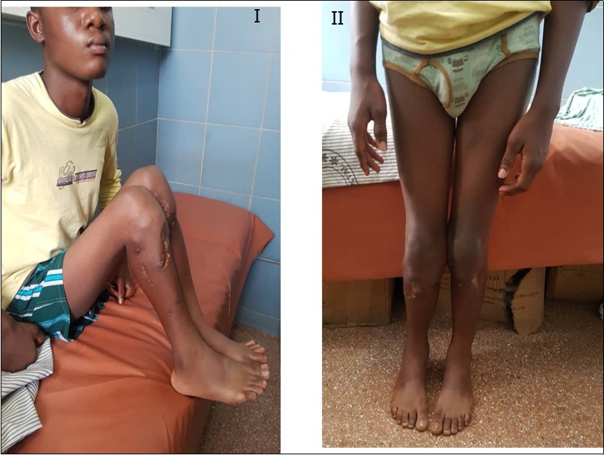
The patient is ambulatory and now has painless knees, a remarkable improvement in gait, no lateral knee thrust, no limb length discrepancy and is able to participate in activities at a higher level and is cosmetically optimised. Figure 8.
Figure 8.X-ray of both knees of the patient anteroposterior view, I, and lateral view II, 2yrs post operatively showing correction of the bilateral tibia vara with 8-plate and screws in situ.
Conclusion
We have presented our management of a boy with a late-presenting infantile tibia vara langenskiold type VI using a single-stage double osteotomy of the tibia and fibular-shortening osteotomy. The child had knee pain, abnormal gait and a cosmetic stigma which limited his social integration and activities of daily living prior to surgery. Our surgical intervention has restored him to the company of his peers by successfully correcting his limb deformities. He now ambulates without knee pain, has a remarkable improvement in his gait, no lateral knee thrust, no limb length discrepancy and is able to participate in activities at a higher level and is cosmetically optimised.
The single-stage double osteotomy surgical technique for correcting neglected tibia vara and Taylor Spatial Frame stabilization of the osteotomy are a feasible method in a low-resource setting. It is a safe and effective method of correction of the disease and has a favourable 2-year outcome. The child would be followed until skeletal maturity for recurrence due to unpredictable proximal tibial physeal growth.
Financial Support and Sponsorship
Nil
References
- 1.Mayer S W, Hubbard E, Sun D, Lark R, Fitch R. (2019) Gradual Deformity Correction in Blount Disease. J Pediatr Orthop. May/jun.
- 2.Huyssteen van A. (2005) L.Double-elevating osteotomy for late-presenting infantile Blount’s disease.The. , Bone & Joint Journal
- 3.N De Sanctis, Della Corte S, Pempinello C, G Di, Gambardella A. (1995) Infantile type of Blount's disease: Considerations concerning etiopathogenesis and treatment. , J Pediatr Orthop B 4, 200-203.
- 6.Cook S D, Lavernia C J, Burke S W, Skinner H B, Haddad RJ Jr. (1983) A biomechanical analysis of the etiology of tibia vara. , J Pediatr Orthop 3, 449-54.
- 7.Arkin A M, Katz J F. (1956) The effects of pressure on epiphyseal growth; the mechanism of plasticity of growing bone. , J Bone Joint Surg Am 38, 1056-76.
- 8.Louis S, David W, Selvadurai N. (2010) . Apley’s System of Orthopaedics and Fractures, 9thed , Hodder Arnold, London, UK .
- 9.Carter J R, Leeson M C, Thompson G H, Kalamchi A, Kelly C M et al. (1988) Late-onset tibia vara: A histopathologic analysis. A comparative evaluation with infantile tibia vara and slipped capital femoral epiphysis. , J Pediatr Orthop 8, 187-95.
- 10.Ingvarsson T, Hägglund G, Ramgren B, Jonsson K, Zayer M. (1998) Long-term results after infantile Blount's disease. , J Pediatr Orthop B 7, 226-9.
- 11.Elhanan Bar-On.Treatment of severe early onset Blount’s disease by an intra-articular and a metaphyseal osteotomy using the Taylor Spatial Frame. , Journal of Children's Orthopaedics,2008
- 12.Nirav K.Pandya.Correction of Blount’s disease by a multi-axial external fixation system. , Journal of Children's Orthopaedics,2009
- 13.Langenskiöld A. (1981) Tibia vara: Osteochondrosis deformans tibiae. Blount's disease. Clin Orthop Relat Res. 158, 77-82.
- 14.Hollman F, Korpisah J, A H Ismail, Rompa P, Moh P et al.Staal H M.(2016).W/M serrated osteotomy for infantile Blount's disease in Ghana: Short-term results. , Niger J Clin Pract.19: 443-8.
- 15.La Mont L, McIntosh A, Jo C, Birch J, Johnston C. (2019) Recurrence After Surgical Intervention for Infantile Tibia Vara: Assessment of a New Modified Classification. , Journal of Pediatric Orthopaedics.FEBRUARY 39(2), 65-70.
- 16.Mark.Eidelman.The use of the Taylor spatial frame in adolescent Blount’s disease: is fibular osteotomy necessary?. , Journal of Children's Orthopaedics,2008
- 17.Gkiokas A, Brilakis E. (2012) Management of neglected Blount disease using double corrective tibia osteotomy and medial plateau elevation.https://doi.org/10.1007/s11832-012-0443-x: 1.
- 18.Vukasnović Z. (2013) Treatment of infantile tibia vara - 18-year follow-up: A case report. Srp Arh Celok Lek. May-June.
- 19.Cherkashin A, Samchukov M, Birch J, Da Cunha A.Evaluation of complications of treatment of severe Blount’s disease by circular external fixation using a novel classification scheme. , Journal of Pediatric Orthopaedics B.MARCH 24(2), 123-130.
- 20.Allison S.Treatment of Infantile Blount Disease with Lateral Tension Band Plating. , Journal of Paediatric Orthopaedics.Jan-Feb 32(1), 29-34.
- 21.Masrouha K Z, Sraj S, Lakkis S, Saghieh S. (2011) High tibial osteotomy in young adults with constitutional tibia vara. Knee Surg Sports Traumatol Arthrosc. 19, 89-93.
- 22.Laville J M, Wiart Y, Salmeron F. (2010) Can Blount's disease heal spontaneously? Orthop Traumatol Surg Res. 96, 531-5.
- 23.Li Shuai, Karatzoglou A, Gentile C. (2016) Collaborative Filtering Bandits. Proceedings of the 39thInternational Conference on Research and Development in Information Retrieval , Pisa, Tuscany, IT (SIGIR) 539-548.