
Abstract
Background and Purpose
Providing health care is the basic right of people(1).
Diagnostic radiology is one of the main procedures in health care services and proper benefiting from this technology is brought only under well planning and management(1).
Supervision of the available condition and its comparison with the recommended standards is a key role in assessing assurance from the benefit of these instruments (2). Data show that more than 80% of patients referring to these hospitals need radiology image (3).Improper service causes repetition of radiography and even wrong diagnosis, as a results threatening health of the patients (3) lack of protective barrier leads to the exposure of the staff to X-ray which is obviously carcinogen us (4). It happens that the instruments are not working properly, like of symmetry in X-ray field, defects in collimators, lack of adjusting ray field and X-ray, low quality or defective developing machine, lack of proper protective barrier, using low quality film and drugs, lack of protective barrier for children, all of which cause severe hazards for the patients and staff (4).
Materials and Methods
The crucial aim of medical services is to provide the public with their needs which are very important. The sensitivity of such services is to such an extent that in case of lack of care, the hazards are too high. In evaluation of health services, the first thing is to evaluate the device used. Methods, efficiency, profits and their combination for prevention and eradication of diseases are also important. Therefore to gain this goal, it is necessary the obtain results comparable with recommended standards. The purpose of this study was to access the conditions of radiology units at Mazandaran University hospitals and compare them with the standards of ICRU NCRP and ICRP. Radiology unit is the most expensive section of any hospital for its instruments, manpower and space provided. In a study conducted in 51centers on radiology staff, radiography room and protective barrier, ray leakage, the outcome were 89%, 82%, 77% and 37% respectively. It was found that the condition of these centers regarding the protective barriers is very unsuitable due to unawareness of the leakage (5, 6).Considering the mentioned necessities, in this study, the condition of radiography centers affiliated to the Mazandaran University of Medical Sciences was studied for the type and the rate of problem, in order to provide a proper solving method.
Results
Data were collected through, observation, interviewing and filling questionnaire. Results show that, the situations of the radiology units are for from international standard, to such an extent that it is matched clout 50%.
Conclusion
The results showed that, none of the dark rooms are standard, and do not have proper alarm signal. In 63% of these units there no tiling system about staff protection from radiation.
Defects in radiography room, protective barrier and lack looking rays were 60%, 51% and 47% respectively. Referring to the obtained data, periodic supervision, and obeying of the standards are necessary.
Author Contributions
Academic Editor: Sasho Stoleski, Institute of Occupational Health of R. Macedonia, WHO CC and Ga2len CC, Macedonia.
Checked for plagiarism: Yes
Review by: Single-blind
Copyright © 2021 Vajiheh Keshavarz, et al.
 This is an open-access article distributed under the terms of the Creative Commons Attribution License, which permits unrestricted use, distribution, and reproduction in any medium, provided the original author and source are credited.
Competing interests
The authors have declared that no competing interests exist.
Citation:
Introduction
Providing health care of is the basic right of people 1, 2. Diagnostic radiology is one of the main procedure in health care service the proper benefiting from this technology is brought only under well planning and management 3.
Supervision of the available condition and its comparison with the recommended standards is a key role in assessing assurance from the benefit of these instruments 4. Radiology units of the each hospital 3. Data show that more than 80% of patients referring to hospital need radiology image. 5.
Improper service causes repetition and even wrong diagnosis, as a results threatening health of the patients 6 lack of protective barrier leads to the exposure of the staff to X- ray which is carcinogen 7.
It happens that the instruments are not working properly, like of symmetry in x ray field, defects in collimators, lack of adjusting ray field and x ray, also disorder in developing machine, lack of proper protective barrier, using low quality film and using drugs lack of protective barrier for children, all of which cause severe hazardous for the patients and staff 8.
In a study in USA, it was shown that one of the reasons of unnecessary receiving ray in patients is using higher than normal range of x-ray 4. In a quality control program which was done in different cent radiology center of Iran by Iran atomic organization in cooperation with intonation atomic energy agency , it was found that after execution of quality control program, doses received of the staff and patients reduced to 70% and concomitantly, quality of the radiology film improved to a high extent 9.
In a study performed in 8 mammography center of Tehran University of medical sciences, quality and contrast focusing image max kv with the real , function of photo timer , reproduction of radiation factor, contact of screen film, HVL, temperature function of developing chemicals, sensitivity, dosimeter, control of dark room bulb and environment were studied 10, 11.
It was clear that, the entire center has non standard dark rooms and 87.5 of them have one or more defects in mammography instrument, 37.5% had impermissible mean dose of absorption.
In a study conducted in 51centers to radiology staff, radiography room, protective barrier, ray leakage was 89%, 82%, 77% and 37% respectively it was found that the conditions of such centers for the view point of protective barriers. Is very unsuitable due to unawareness of the available lockage 12, 13.
Considering the mentioned necessities, in this study, the condition of radiography centers affiliated to the Mazandaran University of Medical Sciences was studied for the type and the rate of problem, in order to provide a proper solving method 14, 15.
Materials and Methods
In this study the conditions of the radiography units were compared with the standards. The variables under study were categorized in 6 groups: Radiography room for space, light ray lockage, height and condition and piece of pass cast ventilation, entry door, alarming pester, ray signal, preserving devices, loud speaker, minimum distance of tube from control room and patient's lavatory condition. Condition of radiography instruments like, model and installation date, function of mA. KV. Key and time of key moving in different direction of tubes. Bottoms of instrument rotation, film tray, key function, condition of tube arm and screen copy condition.
Condition of control room for, size , situation towards radiography room light , size of lead glass, height of the lead glass from the floor , situation of lead glass towards x-ray room and control room hygiene . Condition of dark room for entry door , space of the place and distance to radiography room light leakage , internal decoration for reflection of light, ventilation ( power and light resistant) bulb ( type lf filter distance from film, bulb power) , film box (earth wire), charge of drug, needed light, developing device (type and duration of usage) of developing device condition of rollers , condition of installation, position of instrument, blank films and drugs storage for light , ventilation and humidity
Condition of the other facilities such as patients preparing room, waiting room hygiene, staff room (space, facilities and hygiene) staff's lavatory (place, hygiene and space) Hilling room and personal, classification of films.
In this study radiography units were visited and data were collected using questionnaire comprising questions based on radiology standards and protection against ray as well as interview and observation. Detective dose meter FJI model capable of detecting x and gamma rays in the range of 50 kev- 103 keV with energy response of 40-80% was used. An accurate thermometer with measuring range -10C to 150C and accuracy of +1C was used for the determination of developing chemicals and films temperature.
A meter was used to measure the space of radiography and control rooms, height of pass cast, size of lead glass, and height of floor to ceiling. Leakage of ray from door when closed was noticed and lead covering of the wall for protection was considered.
Findings
The obtained results can be divided in two main sections: data from the questionnaire and observation and examinations
Total number of available instruments in the hospitals under study was 50, of which 5 were out of order; they were in use for 1 so 30 years mean duration of 10 years. Results showed that only in 34.4% of the cases there standard see Figure 1.
Figure 1.The condition of radiography room considering the parameters under study in the Mazandaran University of Medical Sciences hospitals.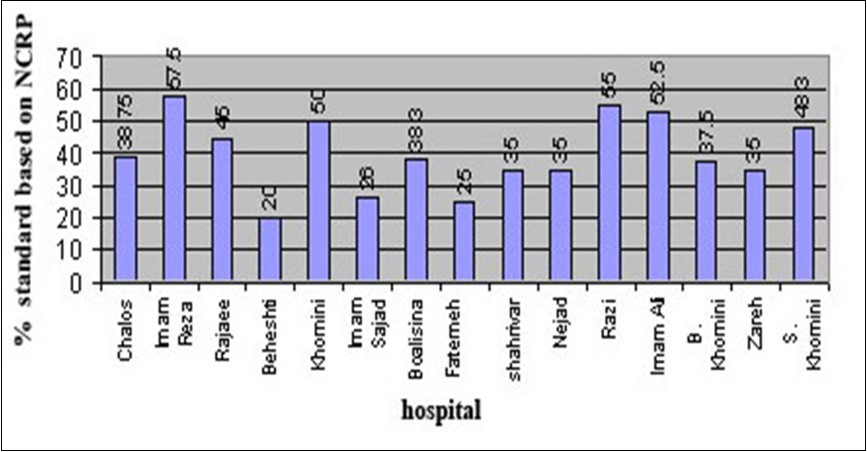
Study of the radiographic instruments through observation and examination showed that their function as compare to the standard criteria is 80.4%. Figure 2 & Figure 3.
Figure 2.Condition of radiography instruments considering the parameter under study in the Mazandaran University of Medical Sciences hospitals.
Figure 3.Condition of control room at the radiography units considering the parameter under study in the Mazandaran University of Medical Sciences hospitals.
In study of dark room condition through direct observation and examination showed that. As compare to the standard criteria 48.82% is standard Figure 4.
Figure 4.Conditions dark room in the radiography unit considering the parameters under study in the Mazandaran University of Medical Sciences hospitals.
In evaluation of the dosimeter, needed protective barriers and results of dosimeter through direct observation data showed that, about 50.4% standard Figure 5.
Figure 5.Condition of dosimeter and protection of staff in the radiographic units, considering the parameter under study in the Mazandaran University of Medical Sciences hospitals.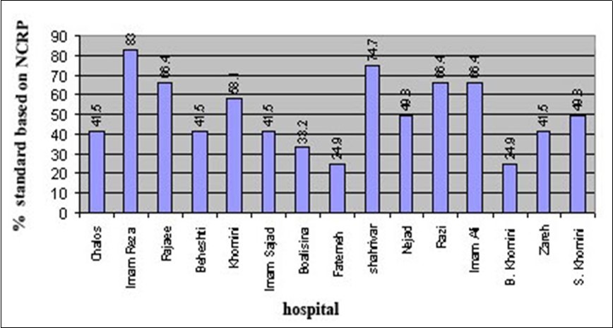
In all radiographic units certain fore seeing must be done for the case preparation of the patients, staffroom, film and chemical storage hilling room and waiting halls. Data showed that the conditions of the adjacent rooms 37.15% is standard Figure 6.
Figure 6.The condition of adjacent rooms in the radiographic units considering the needle parameter under study in the Mazandaran University of Medical Sciences hospitals. (Preparation room waiting room staff room, lavatory filing film and chemical storage).
Data indication that, none of the hospitals are supervised regularly, to such as extent that, some of the defects were unknown lit hill the time of this study, and due to unawareness from the consequents they did not feel hazardous.
Discussion
It was found that 76% of the radiology units had direct screen copy problem, only 96% had thyroid shield –gonad shield and, lead spectacle and lead cover. Considering the significance of such devices to protect children and adolescents against ray, such condition is very disappointing 16, 17. Regarding the study on the presence of alarming signals , some of the units lack poster of irradiation and warning poster for pregnant women and only 40% of the units were in good condition in this regard 18, 19.
About the control of irradiation for staff it was found that 51% of the units had no medical filing system and periodic examination for the staff. Meanwhile 15% of them did not have person in charge of physic health to supervise and follow issues related to the personal protection and periodic check ups 20, 21. Investigation showed that 47% of the units under study showed ray leakage, which demands a serious and prompt intervention. It is worthy to mention that, approximately all of such units lack preservative device and required hard ware 22, 23. All of the units under study had pass cast , but it was found that 67% of them were not efficient and most of the dark rooms had evident leakage of light , and did not have bulb, in 48% the temperature of developing negative film was not suitable 24, 25. The overall score for the units regarding alarming signals in dark room , protection against irradiation , radiology space, protective shield, results of dosimeter and efficiency of different instruments were 50%, 50%, 50%, 40%, 51% and 51% respectively 18. Each staff could take 13 images per day during the first 6 months of 2002.
Generally, considering the results obtained from this evaluation and the repairable defects, regular periodic supervision is absolutely necessary (once every six months) by the expert in order to have better usage of the instruments and facilities
References
- 2. (1977) . International Commission on Radiaton Protection Recommendations of the International Commission on Radiological Protectin. Oxford: ICRP Publication; 26.
- 3. (1981) American Association of Physicists in Medicine. Basic Quality Control in Diagnostic Radiology. AAPM Report. 4.
- 5.Keane B E, Tikhonov K B.. Manual of Radiation Protection in Hospitals and General Practice. WHO 3, 197.
- 6.Mould R F. Department of Medical Physics (1985) Radiation protection in hospitals. , London: Westminster Hospital;
- 8.Rehani M M, Arun Kumar LS, Berry M. (1992) Quality assurance in diagnostic radiology; lnd 1 Radiol Imag. 43-119.
- 9.Moores B M, Stieve F E, Eriskat H. (1989) Technical and Physical Parameters for Quality Assurance in Medical Diagnostic Radiology Tolerances, Limiting Values and Appropriate Measuring Methods. London: British Institute of Radiology. , Report 18.
- 10. (1981) Kodak Image quality control; In Fundamentals of Radiographic Photography; Vol I; Hemel Hempstead; Kodak Ltd;.
- 11. (1968) National Council on Radiation Protection. Medical X-ray and Gamma-ray Protection for Energies up to 10 MeV; Equipment Design and Use; NCRP Report;. , No 33.
- 12.Goldman L W. (1979) Effects of film processing variability on patient dose and image quality, Rockville. Food and Drug Administration, Division of Training and Medical Applications, Burea of Radiological Health;.
- 13.Kodak. (1981) Image quality control. In Fundamentals of Radiographic Photography; Kodak Ltd,Hemel Hempstead;. 1.
- 14.Suleiman O H. (1982) A densitometic evaluation offilm- chemistry- processor systems in the state ofNew Jersey;. , Rockville. Md, U.S
- 15.Simone P. (1994) Radiation Protection in the X-ray Department. , Butter worth Heinemann 23(53), 97-115.
- 16.Manton D J, ej Roebuck, Fordham G L. (1988) Building and Extending a Radiology Department. London: Royal Society of Medicine Servine;.
- 18.Fecdel H, Schneider K, Kahn M. (1989) et Specific principles for optimization of image quality and patient exposure in Pediatric diagnostic imaging. InMoores BM, Walt BF, Eriskat H, et al (eds). Optimiztion of Image Quality and Patient Exposure: in Diagnostic Radiology. London; British Institute of Radiology. Report 20
- 19. (1982) Kodak Radiographic quality;ln Fundamentals of Radiographic Photography. , Hemel Hempstead; 3.
- 21.Sohrabi M, S Borhan Azad. (1999) . , Quality Control & Quality Assurance in Mamography, Forth Iranian Medical Physics Congress 120-231.
- 22.Taghizadeh S.quality control for improvement of image and reduction of absorbent dose. in patient in mammography
- 23.Taghizadeh S.Evaluation in rate of observing protective factors cind some effective factors in quality of radiology image.