
Broken Endodontic Instrument Caused Inferior Alveolar Nerve Paraesthesia: A Case Report.
Abstract
A healthy 55-year-old man was referred to the Department of Endodontics, Oral and Dental Healthy Hospital, Eskişehir suffering from pain and paraesthesia in the left lower lip and chin.A panoramic radiograph revealed the presence of broken endodontic instrument beyond the apex of the mandibular left third molar. A cone beam computed tomography (CBCT) examination was undertaken, which revealed that the broken instrument was inside the mandibular canal. Damage to the inferior alveolar nerve (IAN) secondary to extrusion of a broken endodontic instrument was diagnosed. Extraction of the tooth was decided and the patient was prescribed with 1 mg/kg/day prednisone 2 times/day, once-daily regimen, and 150 mg/day pregabalin, two doses per day, monitoring the progress with periodic follow-up visits. One month after the incident, the signs and symptoms were gone. The complete resolution of the paraesthesia and the control of pain achieved in the present case suggest that surgical removal of broken endodontic instrument extruded into the mandibular canal with the use of prednisone and pregabalin is a good option in the management of inferior alveolar nerve injury.
Author Contributions
Academic Editor: Allauddin Siddiqi, Sir Johan Walsh Research Institute, University of Otago
Checked for plagiarism: Yes
Review by: Single-blind
Copyright © 2016 Selcuk M. Ozbek, et al
 This is an open-access article distributed under the terms of the Creative Commons Attribution License, which permits unrestricted use, distribution, and reproduction in any medium, provided the original author and source are credited.
Competing interests
The authors have declared that no competing interests exist.
Citation:
Introduction
Injury to the inferior alveolar nerve (IAN) following endodontic treatment in the posterior mandibular teeth is a relatively infrequent complication resulting in disabling sensory disturbances such as pain, paraesthesia, and dysaesthesia of the lower lip and chin area.1 Paraesthesia is defined as an abnormal sensation with clinical manifestations such as burning, prickling, tingling, numbness, itching or formication, which is not unpleasant.1, 2
The possible etiologic factors for endodontic related paraesthesia are periapical infection and iatrogenic injury to the nerve.2, 3 Overinstrumentation during root canal treatment with manual or rotary instruments allowing overfilling of mandibular molar and premolar is a potential iatrogenic cause of inferior alveolar nerve injury.4, 5
The suggested therapeutic sequence for endodontically related paraesthesia is the control of pain and inflammation and, whenever possible, the surgical elimination of the cause.6, 7 The primary aim of surgical treatment should be the entire removal of the foreign body and protection of the nerve.6, 8
The aim of this paper is to describe a case of labiomandibular paraesthesia due to broken endodontic instrument extrusion into the mandibular canal, and the use of cone-beam computed tomography (CBCT) to aid the diagnosis and treatment.
Case Report
A 55-year-old man was referred to the Department of Endodontics, Oral and Dental Healthy Hospital, Eskişehir, Turkey, with a chief complaint of pain and paraesthesia in the left lower lip and chin. Medical histories showed no significant information and physical (extra- and intraoral) examination was within normal limits except for the paraesthesia.
The patient reported that 1 week earlier he had endodontic treatment initiated by a general dentist in his left mandibular third-molar. Two days after the beginning of root canal treatment he developed severe pain and paraesthesia in the left lower lip and chin. On reporting this, a panoramic radiograph had been taken by the referring dentist. It showed a broken endodontic instrument located in the apices of the left mandibular third-molar with an extension toward the IAN (Figure 1). Amoxicillin 1g every 12 hours and paracetamol 500 mg every 12 hours was prescribed by his dentist. The prescribed analgesic reduced pain but paraesthesia in the left lower lip and chin continued and prompted referral by his dentist.
Figure 1.Panoramic radiograph of broken instrument located in the apices with an extension toward mandibular canal.
Clinical examinations using a dental probe showed paraesthesia extending from the mandibular midline to the left second premolar both intraorally and extraorally Figure 2. A temporary restoration placed in the occlusal surface of the tooth was observed.
Figure 2.Area of paraesthesia on the first visit.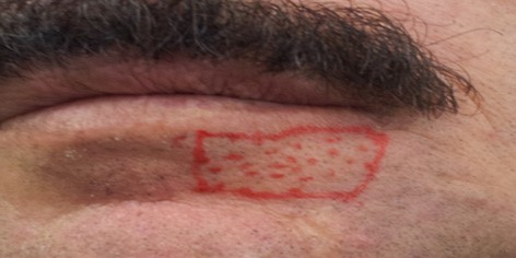
The patient’s symptoms and the close proximity of the broken instrument to the mandibular canal seen in panoramic radiography suggested that the broken endodontic instrument was in contact with the IAN. To confirm this diagnosis, CBCT examination was undertaken, and clearly showed the broken instrument in the periapical area of the mandibular left third molar and that the instrument was inside the mandibular canal. The result was the same as the panoramic examination, broken instrument was noted in the left mandibular canal Figure 3a, Figure 3b.
Figure 3a.CBCT surface examination broken endodontic instrument lodged in the mandibular canal.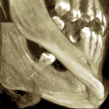
Figure 3b.CBCT examination broken endodontic instrument lodged in the mandibular canal.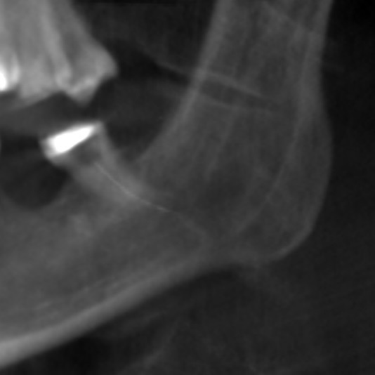
After discussing treatment options the patient refused endodontic treatment and extraction of the lower left third molar was therefore approved. Local anaesthesia was administered in the form of inferior alveolar nerve block. The temporary restoration was removed. The instrument was located and extricated from the canal with the help of fine mosquito forceps and then the tooth was extracted (Figure 4). Panoramic radiograph confirmed satisfactory removal of the broken endodontic instrument from the mandibular canal (Figure 5). The same treatment protocols reported by López-López et al., 20125 were used: the patient was prescribed with 1 mg kg -1 per day prednisone (Dacortin®,30 mg) in two doses, in a gradually reducing regimen on a daily basis, and 150 mg per day pregabalin (Lyrica®, 75 mg; Pfizer SL, Barcelona, Spain), two doses by day, monitoring the progress with periodic follow-up visits.
Figure 4.Retrieved broken endodontic instrument.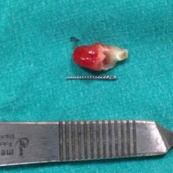
Figure 5.Panoramic radiographs confirmed removal of the broken endodontic instrument from the mandibular canal.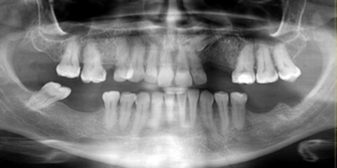
Five days later, the patient had no pain, and paraesthesia in the region of the left lower lip and chain had decreased. Three weeks later, the paraesthesia was significantly reduced and pregabalin treatment was completed. The patient reported a gradual reduction in paraesthesia over the following ten days, and one month after the incident, the signs and symptoms were gone. The patient was satisfied with the outcome and refused to undertake any further radiographic or CBCT examination of the area.
Discussion
Iatrogenic injury following oral surgical procedures or inadequate dental treatment is the most frequent cause of sensory disturbances in the distribution of the inferior alveolar and mental nerve.1 In endodontics, one of the potential iatrogenic causes of this problem is the incorrect treatment of the root canals (overextension and/or overfilling) of a lower molar or premolar.9 Preventive measures such as the use of an electronic apex locater and the application of a good apical stop, will help avoid endodontic complications such as those caused by broken endodontic instruments.1, 5 The proximity of the mandibular canal to the apices of the premolar and molar teeth requires a careful radiographic diagnosis when endodontic treatment of these teeth is planned. An initial pretreatment radiograph of the mandibular teeth will reveal the proximity of the canal to the apices.10 In the present patient, paraesthesia in the left lower lip and chin was caused by the broken endodontic instrument irritating the inferior alveolar nerve sheath. This injury classified as Sunderland’s first degree of nerve injury.11
Studies have indicated that recovery from nerve damage depends on the duration of the trauma and severity of the lesion.1, 12 Reported cases of chemical or thermal injuries showed no recovery from the dysaesthesia or paraesthesia despite the removal of the offending foreign bodies.13 In the case discussed here, the patient recovered from paraesthesia after extracting both the instrument and tooth without any deficits within one month. Along similar lines, Nayak et al., 201113 reported after the surgery named simple deroofing technique the patient complete recovered from her nerve injury within four months.
CBCT scans produce reconstructed multiplanar images that allow the clinician to assess the area of interest three-dimensionally.14 This has been shown to result in more accurate diagnoses compared with intraoral radiographs, which should improve patient management.15, 16 The captured CBCT data can reveal additional relevant information about neighbouring anatomical structures (e.g. mandibular nerve). According to American Association of Endodontists (AAE) and the American Academy of Oral and Maxillofacial Radiology (AAOMR) guidelines, the use of CBCT should be prescribed only after weighing the risks of radiation exposure with the benefit of the diagnostic information that can be obtained from the scan.17 In this case the use of CBCT was justified and identified the exact position of the broken instrument in relation to the mandibular nerve canal.
Treatment options for decompression of the nerve can include extraction of the tooth and cleaning the socket, apicotomy of the overfilled tooth with cleaning around bone, and decortication of the mandible achieved laterally through intraoral approach.18 In the present case, the patient refused endodontic treatment, and decided to extract the third lower left molar. Therefore, extraction was agreed upon including anti-inflammatory treatment with prednisone and analgesic treatment with pregabalin.
Except for duloxetine and pregabalin medications most of the pain treatments used for neuropathic pain have not been approved by FDA.7, 19 Pregabalin has shown analgesic activity in preclinical and clinical models, and appears to have significant analgesic properties following following third molar extraction.7, 20, 21 Pregabalin is a safe and well-tolerated new treatment for neuropathic pain.5, 22 Taking into account that broken endodontic instrument extrusion into the mandibular canal damages the inferior alveolar nerve triggering neuropatic pain, the use of pregabalin in the case reported here was justified.
This present case shows that CBCT images may provide a more accurate diagnosis when endodontic-related inferior alveolar nerve paraesthesia is suspected. The complete resolution of the paraesthesia and the control of pain achieved in the present case suggest that surgical removal of broken endodontic instrument extruded into the mandibular canal with the use of prednisone and pregabalin is a good option in the management of inferior alveolar nerve injury.
This case demonstrates destruction of the apical foramen caused by over preparation which may lead to direct physical injury of the alveolar nerve. Great care must be taken when performing root canal therapy, particularly when the root apices are in close proximity to anatomic structures such as the inferior alveolar canal nerve.
References
- 1.Scolozzi P, Lombardi T, Jaques B.Successful inferior alveolar nerve decompression for dysaesthesia following endodontic treatment: report of 4 cases treated by mandibular sagittal osteotomy.Oral Surgery Oral Medicine Oral Pathology Oral Radiology and Endodontics2004;97:. 625-631.
- 2.Andrabi S M, Alam S, Zia A, Khan M H, Kumar A.Mental nerve paraesthesia secondary to initiation of endodontic therapy: a case report.Restorative Dentistry and. Endodontics2014;39: 215-219.
- 3.Knowles K I, Jergenson M A, Howard J H.Paraesthesia associated with endodontic treatment of mandibular premolars.Journal of Endodontics2003;29:. 768-770.
- 4.Escoda-Francoli J, Canalda-Sahli C, Soler A, Figueiredo R, Gay-Escoda C.Inferior alveolar nerve damage because of overextended endodontic material: a problem of sealer cement biocompatibility?Journal of Endodontics2007;33:. 1484-1489.
- 5.López-López J, Estrugo-Devesa A, Jané-Salas E, Segura-Egea J J.Inferior alveolar nerve injury resulting from overextension of an endodontic sealer: non-surgical management using the GABA analogue pregabalin.International Endodontic. Journal2012;45: 98-104.
- 6.Pogrel M A.Damage to the inferior alveolar nerve as the result of root canal therapy.Journal of the American Dental Association2007;138:. 65-69.
- 7.Alonso-Ezpeleta O, Martín P J, López-López J, Castellanos-Cosano L, Martín-González J et al.Pregabalin in the treatment of inferior alveolar nerve paraesthesia following overfilling of endodontic sealer.Journal of Clinical and Experimental Dentistry2014;6:e197-202.
- 8.Grötz K A, Al-Nawas B, de Aguiar EG, Schulz A, Wagner W.Treatment of injuries to the inferior alveolar nerve after endodontic procedures.Clinical Oral. Investigations1998;2: 73-76.
- 9.González-Martín M, Torres-Lagares D, Gutiérrez-Pérez J L, Segura-Egea J J.Inferior alveolar nerve paraesthesia after overfilling of endodontic sealer into the mandibular canal.Journal of Endodontics2010;36:. 1419-1421.
- 10.Poveda R, Bagán J V, Fernández J M, Sanchis J M.Mental nerve paraesthesia associated with endodontic paste within the mandibular canal: report of a case.Oral Surgery Oral Medicine Oral Pathology Oral Radiology and Endodontics2006;102:e46-49.
- 11.Campbell W W.Evaluation and management of peripheral nerve injury.Clinical. Neurophysiology2008;119: 1951-1965.
- 12.Grant G A, Goodkin R, Kliot M.Evaluation and surgical management of peripheral nerve problems.Neurosurgery1999;4:. 825-839.
- 13.Nayak R N, Hiremath S, Shaikh S, Nayak A R.Dysaesthesia with pain due to a broken endodontic instrument lodged in the mandibular canal a simple deroofing technique for its retrieval: case report.Oral Surgery Oral Medicine Oral Pathology Oral Radiology and Endodontics2011;111:e48-51.
- 14.Al-Rawi B, Hassan B, Vandenberge B, Jacobs R.Accuracy assessment of three-dimensional surface reconstructions of teeth from cone beam computed tomography scans.Journal of Oral Rehabilitation2010;37:. 352-358.
- 15.Patel S, Dawood A, Ford T P, Whaites E.The potential applications of cone beam computed tomography in the management of endodontic problems.International Endodontic. Journal2007;40: 818-830.
- 16.Ahlowalia M S, Patel S, Anwar H M, Cama G, Austin R S et al.Accuracy of CBCT for volumetric measurement of simulated periapical lesions.International Endodontic. Journal2013;46: 538-546.
- 17.Ball R L, Barbizam J V, Cohenca N.Intraoperative endodontic applications of cone-beam computed tomography.Journal of Endodontics2013;39:. 548-557.
- 18.Brkić A, Gürkan-Köseoğlu B, Olgac V.Surgical approach to iatrogenic complications of endodontic therapy: a report of 2 cases.Oral Surgery Oral Medicine Oral Pathology Oral Radiology and Endodontics2009;107:e50-53.
- 19.Barrett A P, Smith M W.Maxillary nerve involvement in bacterial endocarditis.International. , Journal of Oral and Maxillofacial Surgery1985;43: 816-817.
- 20.Hill C M, Balkenohl M, Thomas D W, Walker R, Mathé H et al.Pregabalin in patients with postoperative dental pain.European. , Journal of Pain2001;5: 119-124.