
Elasticity Profile of Skin, Neuronal, Cardiac, and Skeletal Muscle Cells after Treatment with the Biofield Energy Healing-Based Proprietary Test Formulation
Abstract
The present study aimed to evaluate the effect of the Trivedi Effect®- Biofield Energy Treated/Blessed Test formulation/item (TI) composed of minerals (magnesium, zinc, copper, calcium, selenium, and iron), vitamins (ascorbic acid, pyridoxine HCl, alpha tocopherol, cyanocobalamin, and cholecalciferol), Panax ginseng extract, CBD isolates, and β-carotene on elasticity of skin, heart, muscle, and neuronal cells in the H9C2 (rat cardiomyocytes), C2C12 (mouse myoblast cells), HaCaT (human keratinocytes), and SH-SY5Y (human neuroblastoma cells) cell line in DMEM medium. The test formulation constituents were divided into two parts; one section was defined as untreated test formulation (UT), while the other portion of test formulation received Biofield Energy Healing/Blessing Treatment (BT) by a renowned Biofield Energy Healer, Mr. Mahendra Kumar Trivedi. The test items were treated with Biofield Energy Healing/Blessing Treatment and divided as Biofield Energy Treated/Blessed (BT) and untreated (UT) test items. MTT data showed that the test formulation in various concentrations was found as safe and nontoxic in the tested concentrations with viability range from 73% to 307%. Young’s modulus (YM) is a measure of cell stiffness, a decrease in YM value indicates increase elasticity of the cells and vice-versa. YM in H9C2 cells were decreased by 9.6% and 66.1% in the BT-DMEM + UT-TI group at 0.1 and 1 µg/mL respectively, as compared with untreated test group. However, C2C21 cells showed increased YM by 443.9% at 1 µg/mL in the UT-DMEM + BT-TI group, while 869.6% increased YM in the BT-DMEM + UT-TI group at 1 µg/mL as compared with untreated test group. However, 314% increased YM was reported in the BT-DMEM + BT-TI group at 1 µg/mL as compared with the untreated test group. However, the value of YM was significantly decreased in the HaCaT cell line by 247.7% (at 1 µg/mL), 225.8% (at 0.1 µg/mL), and 97.9% (at 1 µg/mL) in the UT-DMEM + BT-TI, BT-DMEM + UT-TI, and BT-DMEM + BT-TI group respectively, as compared with the untreated group. In addition, YM was significantly decreased in the SH-SY5Y cell line by 92.6%, 18.1%, and 26.6% at 1 µg/mL in the UT-DMEM + BT-TI, BT-DMEM + UT-TI, and BT-DMEM + BT-TI group respectively, as compared with the untreated group. Overall, the results showed the significant decreased YM among the SH-SY5Y, HaCaT, and H9C2 cells, while it was increased in the C2C21 cell line. Thus, the mechanical properties of cells such as cellular function, including shape, motility, differentiation, division, and adhesion to its surrounding extracellular matrix were improved. Overall, it can be useful in many disease progressions with improved cellular elasticity and its associated complications/symptoms.
Author Contributions
Academic Editor: Fuwen Yuan, Duke University, USA.
Checked for plagiarism: Yes
Review by: Single-blind
Copyright © 2021 Mahendra Kumar Trivedi, et al.
 This is an open-access article distributed under the terms of the Creative Commons Attribution License, which permits unrestricted use, distribution, and reproduction in any medium, provided the original author and source are credited.
Competing interests
The authors have declared that no competing interests exist.
Citation:
Introduction
Elastic properties of cells play crucial role in normal physiological processes in living organisms. Cell differentiation is one of such example. In addition, aging of the cells causes changes in the cross linking of extracellular matrix components which further alters the cytoskeletal organization 1, 2. These changes are characterized by alterations in mechanical properties of the cells such as elasticity and rigidity. Elasticity of the cells can be measured by investigating deformability of the cells using techniques such as Atomic Force Microscopy (AFM) 2. AFM is used to determine the Young’s modulus (YM) which is a measure of the stiffness of the biological sample in response to an applied load 1, 3, 4. Cell and tissue stiffness (elasticity) are considered as an important characteristic of normal and diseased cellular states 5. The present study focused on the detection of cellular elasticity using evaluation of YM using H9C2, C2C12, HaCaT, and SH-SY5Y cell lines. Thus, a novel test formulation was designed with the combination of vital minerals (Ca, Zn, Mg, Se, Fe, Cu), vitamins (B12, E, D3, C, B6), and some biological active plant-based extracts such as β-carotene, ginseng, and cannabidiol isolate (CBD). All the minerals and vitamins used in the test formulation have significant functional role to provide vital physiological role 6, 7, 8. Besides, cannabidiol itself has wide range of pharmacological profile and was reported to role in different disorders 9, 10, while ginseng extract is regarded as the one of the best immune booster for overall immunity 11. Vitamins are the major source that builds cellular immunity using different pathways to boost energy level. Vitamin D boosts elasticity of skin, joints, and others parts of body 12, 13. In addition, vitamin D has also been reported to improve arterial stiffness and neuronal plasticity 14. Cannabidiol and Panax ginseng are also known to increase skin elasticity 15, 16, 17. In the present study, effect of Biofield Energy Treatment was determined in cardiac, muscle, neuronal and skin cells.
Novel test formulation was evaluated to study the effect on the level of YM, which in terms represents cellular elasticity. Further, the test formulation and cell line media (DMEM) were treated with Biofield Energy Healing/Blessing Treatment by a renowned Biofield Energy Healer. Biofield Energy Healing Treatment, as a Complementary and Alternative Medicine (CAM) is successfully reported and accepted by National Center for Complementary/Alternative Medicine (NCCAM) against various disorders 18, 19, 20, 21. Various CAM therapies have been accepted by the National Centre of Complementary and Integrative Health (NCCIH) along with the Biofield Energy Healing/Blessing, such as deep breathing, Tai Chi, yoga, therapeutic touch, Reiki, chiropractic/osteopathic manipulation, relaxation techniques, pranic healing, meditation, homeopathy, Ayurvedic medicine, movement therapy, mindfulness, traditional Chinese herbs and medicines in biological systems, etc. 22, 23. However, the Trivedi Effect®-Consciousness Energy Healing/Blessing Treatment to have beneficial impact on various living and non-living things in different disciplines like materials science 24, 25, agriculture science 26, antiaging 27, gut health 28, nutraceuticals 29, pharmaceuticals 30, and overall human health and wellness. In this study, the authors sought to study the impact of the Biofield Energy Treatment (the Trivedi Effect®) on the given novel test formulation on the level of cellular elasticity in H9C2, C2C12, HaCaT, and SH-SY5Y cell lines in terms of YM.
Material and Methods
Chemicals and Reagents
Pyridoxine hydrochloride (vitamin B6), calcitriol, zinc chloride, magnesium (II) gluconate, and β-carotene (retinol, provit A) were purchased from TCI, Japan. Copper chloride, cyanocobalamin (vitamin B12), calcium chloride, vitamin E (Alpha-Tocopherol), cholecalciferol (vitamin D3), iron (II) sulfate, and sodium carboxymethylcellulose (Na-CMC) were procured from Sigma-Aldrich, USA. Ascorbic acid (vitamin C) and sodium selenate were obtained from Alfa Aesar, India. Cannabidiol isolate and panax ginseng extract were obtained from Panacea Phytoextracts, India and Standard Hemp Company, USA, respectively. Imipramine Hydrochloride was purchased from Sigma, USA. H9C2 (rat cardiomyocytes), C2C12 (mouse myoblast cells), HaCaT (human keratinocytes), and SH-SY5Y (human neuroblastoma cells) cell lines were procured from NCCS, Pune. Positive control, calcitriol purchased from TCI Chemicals, However, cell line medium such as DMEM, DMSO, FBS, EDTA, and MTT were procured from Genexlife, Protaq Biomedical, Genexlife, Parshuram and Parshuram traders, respectively.
Cell Culture
H9C2, C2C12, HaCaT, and SH-SY5Y cell lines were used as test system in the present study. The cell lines were maintained in DMEM growth medium for routine culture supplemented with 10% FBS. Growth conditions were maintained as 37°C, 5%CO2, and 95% humidity and subcultured by trypsinisation followed by splitting the cell suspension into fresh flasks and supplementing with fresh cell growth medium. Three days before the start of the experiment (i.e., day-3), the growth medium of near-confluent cells was replaced with fresh phenol-free DMEM, supplemented with 10% charcoal-dextran stripped FBS (CD-FBS) and 1% penicillin-streptomycin 31.
Experimental Design
The experimental groups consisted of cells in baseline control, vehicle control group (0.05% DMSO with Biofield Energy Treated/Blessed and untreated DMEM), positive control group (Calcitriol) and four different experimental test groups. The experimental groups included the combination of the Biofield Energy Treated/Blessed and untreated test formulation/Medium (DMEM). It consisted of four major treatment groups on specified cells with Untreated (UT)-DMEM + UT-Test item (UT-TI), UT- DMEM + Biofield Energy Treated/Blessed test item (BT-TI), BT- DMEM + UT-TI, and BT- DMEM + BT-TI.
Consciousness Energy Healing Strategies
The novel test formulation was consisted of zinc chloride, iron (II) sulfate, copper chloride, vitamin B6, vitamin B12, vitamin D3, sodium selenate, calcium chloride, ascorbic acid, vitamin E, beta carotene, Panax ginseng extract, cannabidiol isolate and magnesium (II) gluconate. Each ingredient of the test formulation was divided into two parts, one part of the test compound was not received any sort of treatment and were defined as the untreated or control sample. The second part of the test formulation was treated with the Trivedi Effect® - Energy of Consciousness Healing/Blessing Treatment (Biofield Energy Treatment) by a renowned Biofield Energy Healer, Mr. Mahendra Kumar Trivedi under laboratory conditions for ~3 minutes in the research laboratory, Dabur Research Foundation, New Delhi, India without touching the samples. After that, the Biofield Energy Treated/Blessed samples was kept in the similar sealed condition and used as per the study plan. In the same manner, the control test formulation group was subjected to “sham” healer for ~3 minutes, under the same laboratory conditions. The “sham” healer did not have any knowledge about the Biofield Energy Treatment/Blessing. The Biofield Energy Treated/Blessed test medium was also taken back to experimental room for further culture methods.
Determination of Non-cytotoxic Concentration
The single cell suspension of H9C2, C2C12, HaCaT, and SH-SY5Y cells were prepared in DMEM with 10% FBS. The cells were counted on a hemocytometer, while the cells were seeded with specific cell density ((5000) cells in H9C2, while 10,000 C2C12, HaCaT, and SH-SY5Y cells/well/180 µL in DMEM + 10% FBS in 96-well plates). The cells were incubated in a CO2 incubator for 48 hours. After 24 hours, medium was removed, and following treatments were given in medium along with the 10% FBS in various experimental groups. After incubation for 24 hours, the effect of the test formulation on cell viability was assessed by 3-(4, 5-dimethylthiazol-2-yl)-2,5-diphenyl tetrazolium bromide (MTT) assay. About 20 µL of 5 mg/mL of MTT was added to all the wells and incubated at 37°C for 3 hours. The cells were centrifuged to obtain the pellet. The supernatant was removed and 150 µL of DMSO was added to all wells to dissolve formazan crystals. Further, all the wells were reported using optical density (OD) values at 540 nm using Synergy HT microplate reader. The effect of the test formulation on viability of cells was determined using equation 1.
% Cell viability = 100 - % Cytotoxicity - (1)
Where; % Cytotoxicity = {(O.D. of Control cells – O.D. of cells treated with test formulation)/ OD of Control cells} *100
Estimation of Cellular Elasticity
The single cell suspension of H9C2, C2C12, HaCaT, and SH-SY5Y cells were prepared in DMEM and 10% FBS using a hemocytometer. The cells were seeded density of 30,000 cells/well/0.5 mL in 48-well plates and incubated in a CO2 incubator for 24 hours and 95% humidity. The cells were centrifuged to obtain the pellet. Supernatants were removed and cells were resuspended in DMEM with 10% FBS. After treatment in all the experimental test groups, the cells were I ncubated in a 5% CO2 incubator for 48 hours, while H9C2 cells were incubated for 72 hours. After 24 hours of incubation, culture supernatants were collected from each well and stored at -20°C until analysis. The cells were fixed using 1% glutaraldehyde for 20 minutes, RT and processed for elasticity measurement by AFM at IISC, Bangalore. Young modulus was calculated from the force/ distance indentation curves generated during AFM analysis using XEI data processing and analysis software.
Statistical Analysis
The data were represented as mean ± standard error of mean (SEM) and subjected to statistical analysis using Sigma-Plot statistical software (Version 11.0). For multiple comparison One-way analysis of variance (ANOVA) followed by post-hoc analysis by Dunnett’s test and for between two groups comparison Student’s t-test was performed. The p≤0.05 was considered as statistically significant.
Results and Discussion
MTT Assay- Non-cytotoxic Effect of the Test Formulation
The cytotoxic effect of the test formulation was evaluated on H9C2, C2C12, HaCaT, and SH-SY5Y cells using MTT assay. The results were compared with respect to defined positive control such as calcitriol. The cells were treated with the test formulation for 48 hours. The effect on viability of cells was determined after 48 hours of treatment by MTT assay (Figure 1). The cells were treated with the test formulation and in various experimental test groups. Calcitriol (PC) demonstrated 164.8%, 110.5%, 77.7%, and 96.4% cell viability at concentration 500 nM in the H9C2, C2C12, HaCaT, and SH-SY5Y cell lines, respectively. In addition, the test formulation resulted more than 73.11% cell viability at the concentration range of 0.01 µg/mL to 10 µg/mL. The results of percentage cell viability range in all the tested cell lines showed the cell viability range of 73% to 307% in different test formulation groups. Overall, the MTT data suggested that the test formulation along with DMEM groups were found safe at all the tested concentrations range up to maximum 10 µg/mL in the tested cell lines.
Figure 1.Effect of the test item on H9C2, C2C12, HaCaT, and SH-SY5Y cell line for cell viability using the MTT assay. UT: Untreated; BT: Biofield Energy Treated/Blessed; TI: Test Item.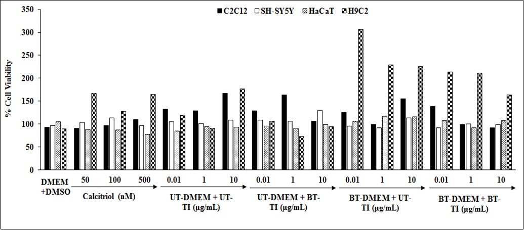
Assessment of Cellular Elasticity in H9C2 Cells
Cellular elasticity in H9C2 cells was determined using percentage change in young’s modulus (YM). The cells were co-treated with the test formulation and stimulated with 0.01 µg/mL to 10 µg/mL for 72 hours. The level of YM was calculated in all the groups, which were compared and presented graphically (Figure 2). The decreased values of YM were calculated on the basis of the untreated test group of DMEM and test formulation. Calcitriol data showed a significant decreased value of YM by 11.1%, 48.3%, and 56.9% at 10, 100, and 500 nM concentrations, respectively. YM in all the treated experimental groups were significantly decreased as compared with the H9C2 cells in DMEM control group, which suggest an improved cellular elasticity of H9C2 cells. Furthermore, the data of the Biofield Energy Treated/untreated test formulation/DMEM combination was compared with respect to the untreated test formulation and DMEM group. Thus, the data suggested that UT-DMEM + BT-TI group showed increase YM by 44.9% and 25.5% in the 0.01 and 0.1 µg/mL, respectively; while decreased YM by 48.8% at 1 µg/mL as compared with the UT-DMEM + UT-TI group. Moreover, 66.1% increased the value of YM was also reported in the BT-DMEM + UT-TI group at 1 µg/mL as compared with untreated test group. Besides, BT-DMEM + BT-TI group showed a increased YM value by 96.8% and 24.9% at 0.01 and 0.1 µg/mL, respectively; while YM value was decreased by 11.5% at 1 µg/mL as compared with the UT-DMEM + UT-TI group. Overall, H9C2 cells (human cardiac muscle cells) treated with Biofield Energy Treated/Blessed medium and TI showed a modulation of elasticity. Modulation in elasticity of cardiac cells (cardiomyocytes) is important for normal heart functions (contractile functions) 32, 33.
Figure 2.Effect of the test item on the level of young’s modulus (YM) in H9C2 cell line after 72 hours. UT: Untreated; BT: Biofield Energy Treated/Blessed; TI: Test Item.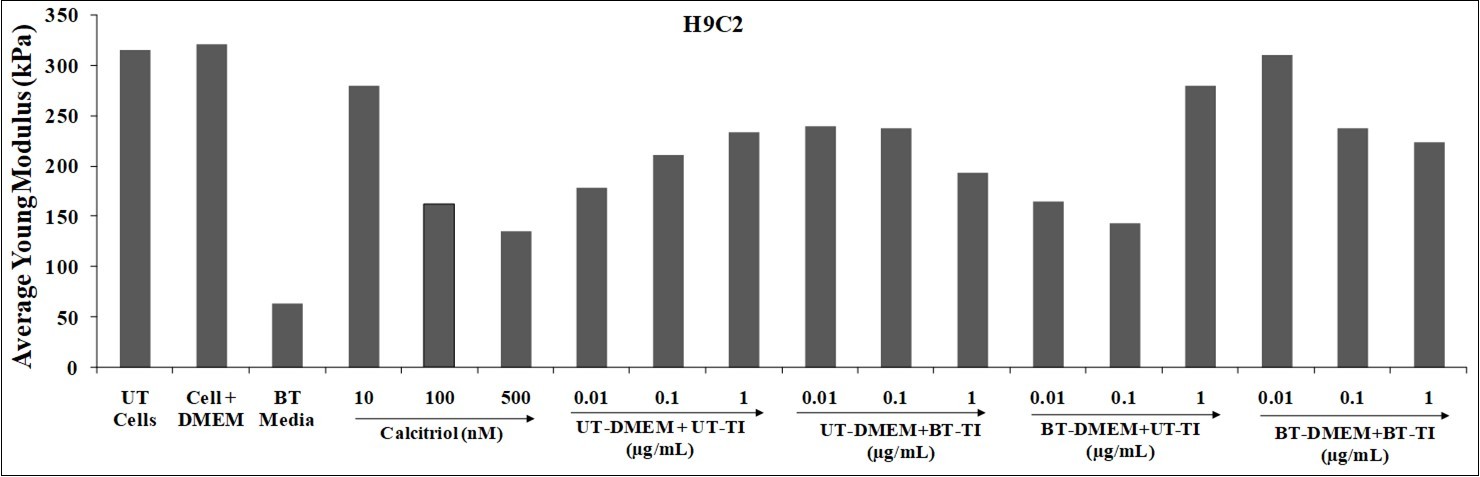
Assessment of Cellular Elasticity in C2C12 Cells
The C2C21 cells were evaluated for cellular elasticity, which was determined using percentage change in young’s modulus (YM) and the data was compared. The cells were co-treated with the test formulation and stimulated with 0.01 µg/mL to 10 µg/mL for 48 hours. The level of YM was calculated in all the groups, which are compared and presented graphically (Figure 3). The increased values of YM were calculated on the basis of untreated test group of DMEM and test formulation. Calcitriol data showed a significant increased the value of YM by 203.8%, 525.3%, and 733.7% at 50, 100, and 500 nM concentrations, respectively as compared with the C2C21 cells in DMEM. The data of Biofield Energy Treated/untreated test formulation/DMEM combination was compared with respect to the untreated test formulation and DMEM group. The increased values of YM were calculated on the basis of untreated test group of DMEM and test formulation. Thus, the data suggested that UT-DMEM + BT-TI group showed decreased value of YM by 103.8% and 84.2% at 0.01 and 0.1 µg/mL, respectively while increased the value of YM by 443.9% at 1 µg/mL as compared with untreated test group (UT-DMEM + UT-TI group). However, 74.1% and 869.6% increased the value of YM was also reported in the BT-DMEM + UT-TI group at 0.1 and 1 µg/mL, respectively as compared with the untreated test group. Besides, BT-DMEM + BT-TI group showed an increased YM value by 314% at 1 µg/mL as compared with the UT-DMEM + UT-TI group. Therefore, in present study increase in YM with Biofield Energy Treatment suggests an increase of differentiation of skeletal muscle cells. Muscle cells have different mechanical properties depending on the physiological processes (cell differentiation, growth, adhesion, etc.). In muscle cells, an increase in YM has been observed which correlates with the extent of differentiation 34. Overall, the present study increase(s) in YM after Biofield Energy Treatment/Blessing suggested increase of differentiation of skeletal muscle cells.
Figure 3.The effect of the test item on the level of young’s modulus (YM) in C2C21 cell line after 48 hours. Vehicle control (DMSO-0.05%), UT: Untreated; BT: Biofield Energy Treated/Blessed; TI: Test Item.
Assessment of Cellular Elasticity in HaCaT Cells
The cellular elasticity in HaCaT cells was determined using percentage change in young’s modulus (YM). The cells were co-treated with the test formulation and stimulated with 0.1 µg/mL to 10 µg/mL for 48 hours. The level of YM was calculated in all the groups, which are compared and presented graphically (Figure 4). The altered values of YM were calculated on the basis of untreated test group of DMEM and test formulation. The values of YM were calculated on the basis of the untreated DMEM and test formulation. Calcitriol data showed a significant decreased value of YM by 7.4%, 28.5%, and 52.7% at 10, 50, and 100 nM concentrations, respectively as compared with the HaCaT cells in DMEM control group. YM in all the treated experimental groups were significantly decreased as compared with the HaCaT cells in DMEM control group, which suggest an improved cellular elasticity of HaCaT cells. Furthermore, the data of Biofield Energy Treated/untreated test formulation/DMEM combination was compared with respect to the untreated test formulation and DMEM group. Thus, the data suggested that UT-DMEM + BT-TI group showed a significant reduced the value of YM by 247.7% at 1 µg/mL as compared with the untreated test group. However, 225.8% and 59.7% decreased the value of YM was also reported in the BT-DMEM + UT-TI group at 0.1 and 1 µg/mL, respectively as compared with the untreated test group. Besides, BT-DMEM + BT-TI group showed a significant decreased YM value by 25.9% and 97.9% at 0.1 and 1 µg/mL, respectively as compared with the UT-DMEM + UT-TI group. Thus, present study concluded that when cells were treated with Biofield Energy Treated/Blessed medium and test formulation, decrease in YM was observed, which indicates an increase in cellular elasticity. In the present study parameter, decrease in YM with Biofield Energy Treatment/Blessing suggests an increase in elasticity of keratinocytes. However, it was reported that modulation in elasticity of skin cells is important for maintenance of skin firmness and elastic properties 35, which was significant improved after Biofield Energy Treatment/Blessing.
Figure 4.The effect of the test item on the level of young’s modulus (YM) in HaCaT cell line after 48 hours. UT: Untreated; BT: Biofield Energy Treated/Blessed; TI: Test Item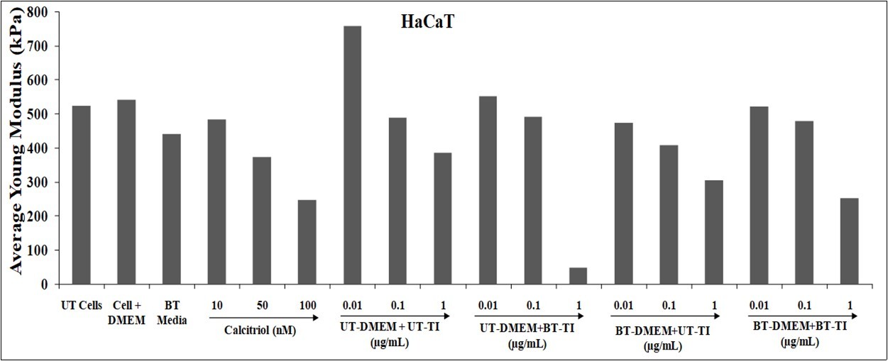
Assessment of Cellular Elasticity in HaCaT Cells
The cellular elasticity in HaCaT cells was determined using percentage change in young’s modulus (YM). The cells were co-treated with the test formulation and stimulated with 0.1 µg/mL to 10 µg/mL for 48 hours. The level of YM was calculated in all the groups, which are compared and presented graphically (Figure 5). The altered values of YM were calculated on the basis of untreated test group of DMEM and test formulation. The values of YM were calculated on the basis of the untreated DMEM and test formulation. Calcitriol data showed a significant decreased value of YM by 7.4%, 28.5%, and 52.7% at 10, 50, and 100 nM concentrations, respectively as compared with the HaCaT cells in DMEM control group. YM in all the treated experimental groups were significantly decreased as compared with the HaCaT cells in DMEM control group, which suggest an improved cellular elasticity of HaCaT cells. Furthermore, the data of Biofield Energy Treated/untreated test formulation/DMEM combination was compared with respect to the untreated test formulation and DMEM group. Thus, the data suggested that UT-DMEM + BT-TI group showed a significant reduced the value of YM by 247.7% at 1 µg/mL as compared with the untreated test group. However, 225.8% and 59.7% decreased the value of YM was also reported in the BT-DMEM + UT-TI group at 0.1 and 1 µg/mL, respectively as compared with the untreated test group. Besides, BT-DMEM + BT-TI group showed a significant decreased YM value by 25.9% and 97.9% at 0.1 and 1 µg/mL, respectively as compared with the UT-DMEM + UT-TI group. Thus, present study concluded that when cells were treated with Biofield Energy Treated/Blessed medium and test formulation, decrease in YM was observed, which indicates an increase in cellular elasticity. In the present study parameter, decrease in YM with Biofield Energy Treatment/Blessing suggests an increase in elasticity of keratinocytes. However, it was reported that modulation in elasticity of skin cells is important for maintenance of skin firmness and elastic properties 36, which was significant improved after Biofield Energy Treatment/Blessing.
Figure 5.The effect of the test item on the level of young’s modulus (YM) in SH-SY5Y cell line after 48 hours. UT: Untreated; BT: Biofield Energy Treated/Blessed; TI: Test Item.
In this research plan, the results showed the significant improved level of cellular elasticity in H9C2, C2C12, HaCaT, and SH-SY5Y cell-lines, which helps in slowdown of the disease progression, disease-related all other symptoms/complications and also reduced the chances of disease susceptibility. This improved cellular differentiation, contractile functions, exonal extensions, and skin elasticity and firmness of the cell lines used in the study after treatment was very significant. Based on the overall data, it suggests that the Biofield Energy Healing/Blessing Therapy was found to be most effective and benefited in order to prevent and protect from the occurrence of any type of diseases. It can also be used as significant way for energy boosting in various disease states that ultimately improve the overall health and quality of life in human.
Conclusions
Cellular elasticity of the cell lines such as H9C2, C2C12, HaCaT, and SH-SY5Y were used and the effect of test formulation was evaluated at different concentrations. Young’s modulus (YM) was calculated using AFM analysis using XEI data processing and analysis software. YM showed a measure of cell stiffness, a decrease in YM value indicates increase elasticity of the cells and vice-versa. MTT analysis for evaluation the non-toxic concentrations were evaluated and data showed that the test formulation in various concentrations was found as safe and nontoxic in the tested concentrations with viability range 73% to 307%. Cellular elasticity using YM was calculated in H9C2 cells, which showed a significant decreased YM values by 9.6% and 66.1% in the BT-DMEM + UT-TI group at 0.1 and 1 µg/mL respectively, as compared with untreated test group. However, C2C21 cells showed increased YM by 443.9%, 869.6%, and 314% at 1 µg/mL in the UT-DMEM + BT-TI, BT-DMEM + UT-TI, and BT-DMEM + BT-TI group, respectively as compared with the untreated test group. Similarly, YM in the HaCaT cell was significantly decreased by 247.7% (at 1 µg/mL), 225.8% (at 0.1 µg/mL), and 97.9% (at 1 µg/mL) in the UT-DMEM + BT-TI, BT-DMEM + UT-TI, and BT-DMEM + BT-TI group respectively, as compared with the untreated group. Furthermore, SH-SY5Y cell line also showed a significantly decreased YM by 92.6%, 18.1%, and 26.6% at 1 µg/mL in the UT-DMEM + BT-TI, BT-DMEM + UT-TI, and BT-DMEM + BT-TI group respectively, as compared with the untreated group. Overall, the Biofield Energy Treated (the Trivedi Effect®) test formulation showed a significant improved cellular elasticity in the tested cell lines, which play a vital role in maintaining various immune and life style related disorders, increase alertness, energy, attention, allergy, Alzheimer, cardiovascular, cancer, diabetes, eye, immune, inflammatory, or Parkinson. Therefore, the Consciousness Energy Healing based test formulation might be suitable alternative nutritional supplement, which could be useful for the management of various immune related disorders. Thus, in conclusion this therapy might also reduce the severity of many acute/chronic diseases and its progression rate.
Acknowledgements
The authors are grateful to Dabur Research Foundation, Trivedi Science, Trivedi Global, Inc., and Trivedi Master Wellness for the assistance and support during the work.
References
- 1.Kuznetsova T G, Starodubtseva M N, Yegorenkov N I, Chizhik S A, Zhdanov R I. (2007) Atomic force microscopy probing of cell elasticity. , Micron 38, 824-833.
- 2.Pasikowska M, Kobiela T, Dulińska M I, Eris I. (2015) The impact of anti-aging peptides on mechanical properties of fibroblasts. Proceedings–23. IFSCC Conference (http://tst.pg2.at/abstracts/data/full_papers/full_paper_134.pdf) , Zurich .
- 3.Kobiela T, Lelen-Kaminska K, Stepulak M, Lekka M, Malejczyk M et al. (2013) The influence of surfactants and hydrolyzed proteins on keratinocytes viability and elasticity. , Skin Res Technol 19, 200.
- 4.Kobiela T, Milner-Krawczyk M, Pasikowska-Piwko M. (2018) The Effect of anti-aging peptides on mechanical and biological properties of HaCaT keratinocytes. , Int J Pept Res Ther 24, 577-587.
- 5.Gavara N. (2017) A beginner’s guide to atomic force microscopy probing for cell mechanics. , Microsc Res Tech 80, 75-84.
- 6.Byrne J H, Voogt M, Turner K M, Eyles D W, McGrath J J et al. (2013) The impact of adult vitamin D deficiency on behaviour and brain function in male Sprague-Dawley rats. , PLoS One 8(8), 71593.
- 9.Peres F F, Lima A C, JEC Hallak, Crippa J A, Silva R H.Abílio VC (2018) Cannabidiol as a promising strategy to treat and prevent movement disorders?. , Front Pharmacol 9, 482.
- 10.Nagarkatti P, Pandey R, Rieder S A, Hegde V L, Nagarkatti M. (2009) Cannabinoids as novel anti-inflammatory drugs. , Future Med Chem 1(7), 1333-1349.
- 11.Kang S, Min H. (2012) Ginseng, the 'Immunity Boost': The effects ofPanax ginsengon immune system. , J Ginseng Res 36(4), 354-368.
- 12.Schagen S K, Zampeli V A, Makrantonaki E, Zouboulis C C. (2012) Discovering the link between nutrition and skin aging. , Dermatoendocrinol 4(3), 298-307.
- 13.Mostafa W Z, Hegazy R A. (2015) Vitamin D and the skin: Focus on a complex relationship: A review. , J Adv Res 6(6), 793-804.
- 14.Dong Y, Stallmann-Jorgensen I S, Pollock N K, Harris R A, Keeton D et al. (2010) A 16-week randomized clinical trial of 2000 international units daily vitamin D3supplementation in black youth: 25-hydroxyvitamin D, adiposity, and arterial stiffness. , J Clin Endocrinol Metab 95(10), 4584-91.
- 15.Song K C, Chang T S, Lee H, Kim J, Park J H et al. (2012) ProcessedPanax ginseng,Sun ginsengincreases Type I collagen by regulating MMP-1 and TIMP-1 expression in human dermal fibroblasts. , J Ginseng Res 36(1), 61-67.
- 16.Hwang E, Park S Y, Yin C S, Kim H T, Kim Y M et al. (2017) Antiaging effects of the mixture ofPanax ginsengandCrataegus pinnatifidain human dermal fibroblasts and healthy human skin. , J Ginseng Res 41(1), 69-77.
- 18.Maizes V, Rakel D, Niemiec C. (2009) Integrative medicine and patient-centered care. , Explore (NY) 5(5), 277-289.
- 19.Bischof M, Del Giudice E. (2013) Communication and the emergence of collective behavior in living organisms: A quantum approach. Mol Biol Int. 987549.
- 20.Cassidy C M. (2004) What does it mean to practice an energy medicine?. , J Altern Complement Med 10(1), 79-81.
- 21.Barnes P M, Bloom B, Nahin R L. (2008) Complementary and alternative medicine use among adults and children: United States. , Natl Health Stat Report 12, 1-23.
- 22.Fan K wai. (2005) National Center for Complementary and Alternative Medicine Website. , J Med Libr Assoc 93, 410-412.
- 23.Wisneski L, Anderson L. (2009) The Scientific Basis of Integrative Medicine. , Boca Raton, FL: 205.
- 24.Trivedi M K, Branton A, Trivedi D, Jana S. (2021) Effect of consciousness energy healing treatment on the metal profile and properties of tellurium. , Eng Technol Open Acc 3(5), 555623.
- 25.Mahendra K T, Alice B, Dahryn T, Snehasis J. (2021) Consciousness energy healing treatment impacted the isotopic abundance ratio of 6-Mercaptopurine (6-MP). Nov Appro Drug Des Dev. 5(5), 555673.
- 26.Trivedi M K, Branton A, Trivedi D, Nayak G, Mondal S C et al. (2015) Morphological characterization, quality, yield and DNA fingerprinting of biofield energy treated alphonso mango (Mangifera indicaL.). , Journal of Food and Nutrition Sciences 3, 245-250.
- 27.Trivedi M K, Jana S. (2021) Anti-aging activity of biofield energy treated novel proprietary test formulation by assessment of vital biomarkers in cerebrospinal fluid (CSF) in Sprague Dawley rats. , On J Neur & Br Disord 5(2).
- 28.Trivedi M K, Jana S. (2021) Evaluation of biofield energy healing treatment based proprietary test formulation on gut health potential in colon cancer cell line (HT-29). , J Pharmacol Clin Res 8(4), 555743.
- 29.Trivedi M K, Branton A, Trivedi D, Jana S. (2021) Isotopic abundance ratio analysis of consciousness energy healing treated folic acid. , Food Nutr Current Res 4(2), 290-295.
- 30.Trivedi M K, Branton A, Trivedi D, Jana S. (2020) The consciousness energy healing treatment and its impact on the isotopic abundance ratio analysis of flutamide. , Drug Des Int Prop Int J 3(5).
- 31.Czekanska E M, Stoddart M J, Richards R G, Hayes J S. (2012) In search of an osteoblast cell model forin vitroresearch. , Eur Cells Mater 24, 1-17.
- 32.Lim C C, Sawyer D B. (2005) Modulation of cardiac function: Titin springs into action. , J Gen Physiol 125(3), 249-252.
- 34.Collinsworth A M. (2002) Apparent elastic modulus and hysteresis of skeletal muscle cells throughout differentiation. , Am J Physiol Cell Physiol 283, 1219-1227.