Communicating Branch of the Mental Nerve and Facial Nerve
Abstract
As peripheral branches of the mandibular nerve, the mental nerve and facial nerve communicate with each other. However, investigations have not always been described in the classic anatomical texts. It remains unknown how nerve fibers of this communicating branch converge at the micro level. Thus, the objective of the present study was to observe in detail the macro and micro levels of the communicating branch of mental and facial nerves. We used five cadavers (10 samples) to conduct experiments in anatomical practice at Tokyo Dental College. A macroscopic observation was made, and the communicating branch of the mental and facial nerves was removed as a single mass. We created serial sections of this branch using the standard method and observed communicating branches of these two nerves under microscopy. As a result, the communicating branch of the mental and facial nerves was completely fused at the perineurium level. It has been reported that the mental nerve includes a small amount of autonomic nerve fiber. As for these findings, similar findings were observed for all 5 bodies and 10 sides. Thus, we believe that autonomic nerve fibers derived from the facial nerve converge with the mental nerve via this communicating branch.
Author Contributions
Academic Editor: Manal ElSawaf, anta University, al-Gaish Street, Tanta, Gharbia, Egypt.
Checked for plagiarism: Yes
Review by: Single-blind
Copyright © 2021 Hidetomo Hirouchi, et al.
 This is an open-access article distributed under the terms of the Creative Commons Attribution License, which permits unrestricted use, distribution, and reproduction in any medium, provided the original author and source are credited.
Competing interests
The authors have declared that no competing interests exist.
Citation:
Introduction
The mental nerve “is a branch of the inferior alveolar nerve that emerges from the mental foramen and immediately branches off into three to four peripheral branches, displaying out in a fan shape and is distributed to the chin and lower lip” 1.
The marginal mandibular branch of the facial nerve is:
“a peripheral branch off of the parotid plexus that emerges from the anterior margin near the inferior corner of the parotid gland, follows a course forward along the lower mandible and communicates with the facial artery on its way. The marginal mandibular branch then continues forward, enters the inferior portion of the depressor anguli oris and innervates muscles such as the inferior half of the orbicularis oris, the depressor anguli oris, the mental nerve, the depressor labii inferioris, and the platysma” 1.
The mental nerve is a sensory nerve, and the marginal mandibular branch of the facial nerve is a motor nerve. Regarding the communicating branch by which these two different nerve types connect, Kamijo 2 has a photograph in his The Anatomy of the Oral Cavity, and Sato and Akita 3 depict the general location using a simple diagram in their Japanese Bodies as “a communicating relation between the peripheral branch of the parotid plexus and the trigeminal nerve,” and describe it in their text as “between the mental nerve and the buccal and marginal mandibular branches.” Other books either include a very minimal information, such as a single line in the text (an example is quoted above) or provide a diagram without any explanation. Most books do not mention the marginal mandibular branch at all. In some cases, this inconspicuous structure was been reported to be a communicating branch and with very close adhesion. However, these reports have also speculated the degree to which the communicating branch is connected to the epineurium, perineurium, and endoneurium on the nerve fiber level. Microscopic observations have been attempted, and the results have led to a consideration of the type of nerve fibers that are present in the communicating branch and the significance of the type of nerve fiber.
Materials and Methods
Five samples (age range: 72 to 86) were obtained from the Department of Anatomy at Tokyo Dental College, and their use in research required approval by the university ethics committee (Yamamoto M, No. 922). The samples consisted of five Japanese adult male cadavers fixed in 10% formalin. Surface dissection was performed using the standard methods. The skin of the face was dissected using scalpel, tweezers, and shears. Next, the fatty tissue, connective tissue, dermal muscles, and associated tissues were removed to allow the vasculature and nerves to maintain their original positions. We were careful to ensure that the natural placements of these structures were preserved despite the loss of supportive tissue. First, we confirmed the location of the mental foramen. Then, as the communicating branch of the mental nerve and facial nerve were identified at the chin and lower lip, we dissected the facial nerve to the anterior margin of the parotid gland to observe the marginal mandibular branch (Figure 1). Next, we removed the wall formed by bone on the buccal side of the mandibular canal to enable observation of the nerves and blood vessels within the buccal canal. Bone removal was performed using a saw and mallet to break up the cortical bone. Using tweezers, we grasped bone fragments and removed them. The inferior alveolar nerve was removed from the mandibular canal to preserve the connectivity between the mental and facial nerves.
Figure 1.Dissection of the region of the mental nerve and the marginal mandibular branch of the facial nerve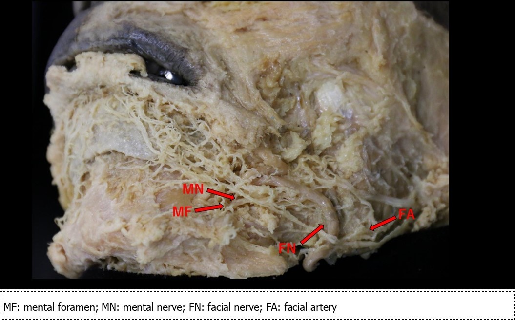
Regarding the region measuring approximately 2 cm from the anterior loop to the peripheral side, the blood vessels and nerves were removed including the confluence of the facial nerve with the inferior alveolar nerve/mental nerve. This was then embedded in paraffin after the removed fat, and series of 30-µm thick sections were prepared. The sections were subjected to hematoxylin and eosin staining, and the courses, confluences, and separations of the nerves were observed under a microscope with the magnification set between ×2 and ×10.
Results
Mental Nerve
The mental nerve (Figure 2) emerges from the mental foramen and advances 5 mm in a posterosuperior direction, at which point the anguli oris branch branches in a superior direction. It then curves in an anterior direction and, at approximately 3 mm, it branches into three. The inferior labial branch moves in an anterosuperior direction to innervate the lower lip. Moving in a distal direction, the first and second of three branches form from the same trunk. On the external side of this common trunk, a branch off the inferior labial branch advances 2 mm, at which point the mental nerve forms a common origin with the inferior labial branch. This mental nerve then turns toward the anteroinferior direction and enters the mentalis muscle.
Figure 2.Sketch of the mental nerve, marginal mandibular branch of the facial nerve and the facial artery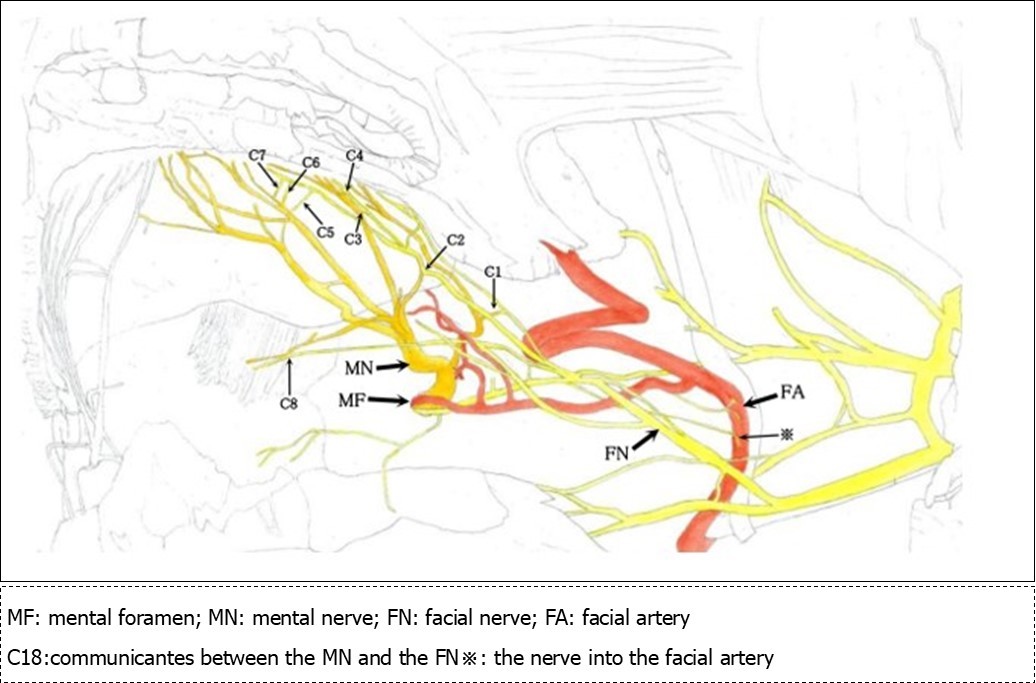
The various branches of the mental nerve belong to a communicating branch that joins with the marginal mandibular branch of the facial muscle. In addition, a relatively thick branch of the marginal mandibular branch of facial muscle communicates with the inferior arterial arch of the alveolar artery and the facial artery at the mental foramen. This characteristic was similar among all specimens.
Marginal Mandibular Branch of the Facial Artery
Two branches extend from the anterior margin of the inferior corner of the parotid gland. The superior branch runs parallel to the mandibular base, while the inferior branch runs anteriorly along the corner of the mandible, after which (and immediately before encountering the facial artery and vein) it branches off into two branches. The first branch follows the inferior margin of the mandible, and the second branch turns in an anterosuperior direction. The latter of these two passes across the facial artery, vein, and mentalis branch of the facial artery and reaches the lower lip. During its course, at the point immediately after passing over the mentalis branch, it fuses with a the buccal branch of the facial nerve, and the branches that arise from this fusion advance toward the lower lip (2 branches) and mentalis (1 branch), enter the mental foramen (1 branch), and a fine branch (1 branch) runs in a posterior direction toward the facial artery. The branch that moves toward the lower lip is a branch that communicates with the anguli oris branch and inferior labial branch (Figure 2, C1-7). The branch that advances toward the mentalis adheres to the mentalis branch of the mental nerve (Figure 2, C8), and the branch that enters the mental foramen adheres to the inferior alveolar nerve immediately after entering the mandibular canal (Figure 3F). There is one fine branch that branches off from the fusion point and advances toward the facial artery (Figure 2※). Prior to the fusion point, there was a branch that branched off the buccal branch and advanced toward the facial artery. The left inferior labial artery was missing, but a similar feature was observed in all specimens.
Figure 3.Mental nerve trunk and facial nerve contiguous with the inferior alveolar nerve removed from the mandibular canal
The location of the Communicating Branches and Adhesions Between the Mental Nerve and Facial Nerve
Communicating branches and adhesions between the mental nerve/inferior alveolar nerve and the marginal mandibular branch of the facial nerve were as follows:
Communication between the anguli oris branch and marginal mandibular branch occurred at a point 1.5 cm from the anguli oris and advanced in a posteroinferior direction at a 45-degree angle (Figure 2, C1)
Communication between the inferior labial branch and marginal mandibular branch at the center of the lower lip, which formed at the most inferior portion of the oral vestibule, which was inferior to the anguli oris (Figure 2, C2)
Communication between the inferior labial branch and the marginal mandibular branch at 1 cm from the vermilion border of the lower lip (Figure 2, C3-7)
Communication between the mental nerve and marginal mandibular nerve inferior to the mentalis muscle (Figure 2, C8)
Communication between the inferior alveolar nerve main trunk and the marginal mandibular branch immediately following entrance into the mandibular canal from the mental foramen (Figure 3, F)
Histological Observations Using the Serial Sections
From the medial side of the facial nerve, we made thin slices that terminated at the anterior loop. These were used to make histological observations of the communicating branches of the facial nerve and mental nerve. At first, we observed the marginal mandibular branch along with blood vessels and connective tissue. There were several nerves, and there were repeated ruptures, fusions, confluences, and separations at the perineurium level, particularly at the epineurium. Once the inferior alveolar nerve appeared in the section (Figure 4A), we first observed that it was connected at the epineurium (Figure 4C). Next, we observed that the nerve fibers formed a confluence immediately following connection with the perineurium (Figure 4D, E). Following this confluence, there was a complete fusion within the perineurium, as it was impossible to differentiate between the two with hematoxylin and eosin staining. I observed similar findings in all specimens.
Figure 4.Fusion between the inferior alveolar nerve and the marginal mandibular branch of the facial nerve as observed microscopically Bar = 200um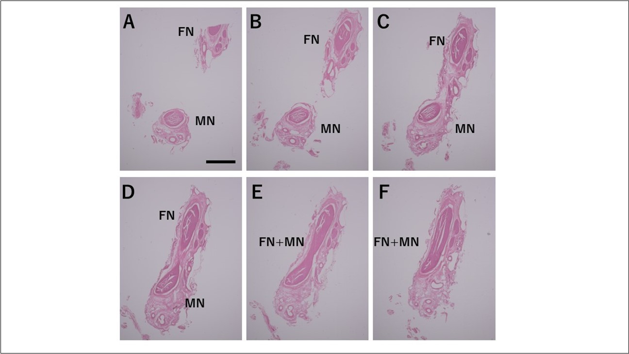
Discussion
Nerve courses in the present case as compared to normal anatomy
The branches of the mental nerve in the present case were as follows: First, there was one independent anguli oris branch. Next, there were three inferior labial branches, the third of which branched from the distal location and was the origin of the mental branch. This origin pattern is the most common and is observed in 28.3% of the Japanese adult population 4. In accordance with the Hosaka study, we assessed the number, courses, and distributions of the various branches as follows 4. There was one anguli oris branch (73.3%) that advanced in an anterosuperior direction after bending in a posterior direction (28.8%) and was distributed to two-thirds of the lower lip from the center (71.6%). There were three inferior labial branches (5.0%) that advanced in an anterosuperior direction after bending in a posterior direction after emerging from the mental foramen (36.6%) and were distributed to one-third of the lower lip from the corner (25.0%). There was one mental branch (56.6%) that originated from the inferior labial branch (58.3%), extended in an anteroinferior direction (1.6%), and was distributed to certain regions of the mentalis (31.6%). The number of inferior labial branches and the course of the mental branch were slightly unusual, but generally speaking, they were consistent with the number of branches and courses observed in most Japanese individuals.
In the present case, the course of the marginal mandibular branch of the facial nerve originated in two branches from the anterior margin of the parotid gland. The superior branch ran parallel to the mandibular base, while the inferior branch split into two branches immediately prior to the facial artery and vein. One of these two branches ran parallel to the mandibular base, while the other bent in a superior direction toward the lower lip and chin. According to Takahashi 5, in 40.1% of cases, there were two main marginal mandibular branches, and in 9.3% of cases they ran a course that is both horizontal and bends upward. Thus, the present case, while not all that exceptional, it was unusual.
What is the normal anatomy for the communicating branches, adhesions, and fusions that are observed for the normal mental nerve and, rarely, between the marginal mandibular branch of the facial nerve? Based on the fact that, in many cases, the total number of branches of the mental nerve is nine branches, each of which originates in a well-balanced manner from one or more locations, the author felt that the present case would not be a particularly unique case. However, without performing multiple dissections using the same methodology, it is impossible to know for sure. Thus, the author consulted the literature and found that multiple studies that reported this same structure 6, 7, 8, 9. All of these were books on the average physiological structure of the human body. The universal nature of the data in these works span a number of years with a substantial amount of data supporting the structural organization described in this report. Nevertheless, major works such as Gray and Netter do not include any such descriptions. Although not mentioned in famous works that incorporate broad information, there are numerous smaller works that include detailed observations mention this particular structural organization. Considering this fact, we believe that they support the conclusions of this paper regarding the network of communicating branches between the mental nerve and the marginal mandibular branch of the facial nerve as a normal structure.
The fact that they are normal structures, and the fact that communicating branches were present in the case we studied, indicates that these structures were present even when the course(s) of one or more nerves was somewhat unusual, and that they did not arise simply due to the fact that the course of the facial nerve was unusual. Still, it remains unknown whether the structural aspects of the branches formed at random without any underlying meaning or whether their precise formation performs some function that should be studied. Determining whether the communication branches arose because they were essential remains to be elucidated by future studies.
The Origin of the Nerves that Compose the CommuniCating Branches and the Types of Nerve Fibers
Based on the results obtained in this study, the mental nerve and the marginal mandibular branch of the facial nerve are completely fused at the perineurium. Most nerve diagrams indicate that the mental nerve is only a sensory nerve, and that the marginal mandibular branch of the facial nerve is a pure motor nerve. However, we found that there is a need for autonomic nerves to be present in the lower face region, where there are salivary glands and sweat glands, including the lower lip and chin (the regions of these nerves). Hence, we aimed to determine where these nerves originate. In the present study, we observed that the vasomotor nerve of the facial artery branched off from the buccal branch and the marginal mandibular branch of the facial nerve (Figure 2※), and we identified the fact that there are sympathetic and parasympathetic fibers in the facial nerve. This was also confirmed micro-anatomically. Figure 5 shows a cross-section of the optic nerve, and Figure 6 shows a cross-section of the mental foramen region of the mandibular nerve. Tyrosine hydroxylase immunostaining stained the sympathetic nerves (provided by lecturer Masahito Yamamoto of the Department of Anatomy, Tokyo Dental College). The trigeminal nerve includes sympathetic fibers, and when the two sections are compared, the optic nerve has a substantial number of such fibers, whereas the mandibular nerve has very few. Matsubayashi 10 wrote extensively on this issue. It is known that the trigeminal nerve, which is the great auricular nerve, auriculotemporal nerve, and zygomatic branch of the facial nerve, consists of cutaneous nerves of the face and sympathetic nerve fibers, although there are few sympathetic nerve fibers in the maxillary nerve or mandibular nerve.
Figure 5.Sympathetic nerve fibers of the optic nerve (anterior facial nerve: TH staining) Bar = 100um
Figure 6.Sympathetic nerve fibers of the mandibular nerve (mental nerve: TH staining) Bar = 100um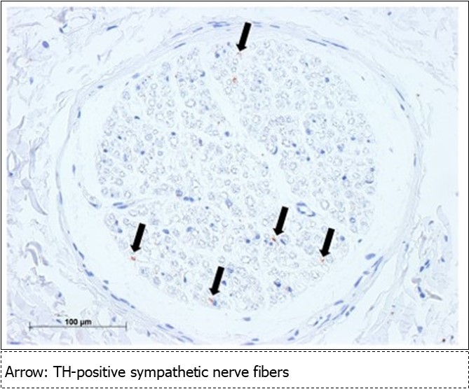
Although it is not completely known which nerve fibers are present in which nerves, the following is a summary of what has been elucidated thus far:
Normally, the mental nerve and facial nerve have intercommunicating branches (Figure 2, all references).
There is a physiological need for sympathetic nerves in the region of the mental nerve.
There are few sympathetic nerves in the mental foramen region of the mandibular nerve (Figure 6, Matusbayashi et al. 10).
There are vasomotor nerves in the marginal mandibular branch of the facial nerve, which includes autonomic nerves (Figure 2)
When the above points are considered in simultaneously, the following hypothesis can be presented:
“The autonomic nerves required for vasomotor control and for the secretions of the glands in the region of the mental nerve are included only in very small amounts in the mental nerve itself, and this deficiency is made up for by communicating branches mainly with the marginal mandibular branch of the facial nerve.”
It is possible to say the same thing regarding the maxillary nerve. To verify this hypothesis, in addition to the factors listed below, it will also be necessary to conduct novel experiments and clinical case reports:
Verify that the communicating branches between the mental nerve and the facial nerve have normal anatomical structures
Verify that the communicating branches include sufficient amounts of autonomic nerves (sympathetic/parasympathetic).
Verify that before and after the communicating branches, the percentage of autonomic nerve fibers included in the mental nerves increase in ratio.
Verify that the mental nerve innervates the inferior labial gland, sweat glands, and blood vessels.
Verify that trauma to the mental nerve and facial nerve leads to drying of the lower lip, lack of perspiration (reduced perspiration) at the lower lip, and abnormalities in the blood distribution using case studies.
If these issues can be verified with a high degree of certainty, the communicating branches of the mental nerve and the facial nerve, which seem to be meaningless structures, can be added as nerve diagrams that serve as the basis for future research and physiological consistency.
Conclusion
The communicating branches of the mental nerve and marginal mandibular branch of the facial nerve are normal anatomical structures that are fused at the perineurium level. These structures may supply autonomic nerves to the mental nerve region from the facial nerve, although further studies are needed to explore and understand this structure and its functions in-depth.
Acknowledgements
We would like to thank Mr. Hio, a student at Tokyo Dental College, who cooperated with the experiment in this study.
References
- 1.Shinohara H, Mataga I, Kageyama I. (2010) Discussion of clinical anatomy of the lingual nerves.doi: 10.2535/ofaj.87.97.Okajimas Folia Anatomica Japonica.87(3),97-102.
- 3.Sato T, Akita K. (2000) Japanese bodies: A discussion of anatomical variations.University of Tokyo Publishing,502-503.
- 4.Hosaka N. (1960) Anatomical study of the mandibular nerve and the mental nerve. Oral Cavity Anatomical Studies.15,279-289.
- 5.Takahashi H. (1957) Local anatomical study of the peripheral branches of the facial nerve. Dental Report.57(7),1-44.
- 6.Kawai T. (1987) Editorial supervision Anatomical records using diagrams Hirosaki University.429 (Fig. B-345)
- 8.Kosugi K, Abe S, Ide Y. (2000) Anatomical study of the course of the mental nerve: 3D observations using silicon injected specimens. Japanese) , Journal of Dentistry and Basic Medicine 42-67.